
Utility of Fundus Fluorescein Angiography and Efficacy of Ranibizumab Intravitreal Therapy in Peripheral Retinal Vascular Abnormalities of Diabetic Patient: A Case Report
Abstract:
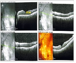
Diabetes predominantly affects the microvascular circulation of the retina. Diabetic retinopathy is a severe complication of diabetes mellitus and a leading cause of diminution of vision. Preventing irreversible vision loss requires early diagnosis of retinal vascular abnormalities. Fundus fluorescein angiography (FFA) is the gold standard for assessing retinal vascular abnormalities and optical coherence tomography confirms the diagnosis. The purpose of this case report was to examine the effectiveness of ranibizumab intravitreal therapy in diabetic patients as well as the usefulness of FFA in evaluating peripheral retinal vascular abnormalities. A 67-year-old adult presented to the emergency department in a tertiary care hospital in Chennai, India, with the sudden onset of painless vision loss in the left eye. Due to irregular medication, he developed diabetic macular oedema. Optical coherence tomography confirmed the presence of macular edema in the left eye and consent was obtained for unilateral pan-retinal photocoagulation (PRP). Despite grid laser, the macular edema persisted and after PRP it was reduced. FFA assisted in treating macular oedema by detecting peripheral retinal ischaemia and administering anti-vascular endothelial growth factor injection (Ranibizumab). The patient's best-corrected visual acuity was improved from 6/36 to 6/9 in the left eye. Research are needed to improve glycaemic management and prompt therapies to help patients with diabetic retinopathy see better and live better lives.
References:
[1].
Raka, M., Daigavhane, S., & Patil, A. A.,
2022, Clinical case report of diabetic retinopathy. Asian J Biomed
Pharmaceut Sci, 12(90),128. https://www.alliedacademies.org/articles/a-clinical-case-report-of-diabetic-retinopathy.pdf
[2]. International
Diabetes Federation, 2019, IDF Diabetes Atlas, 9th ed. Brussels, Belgium, International
Diabetes Federation, https://d-net.idf.org/en/library/542-idf-diabetes-atlas-2019-9th-edition.html
[3]. Suganya, M., &
KalaBarathi, S., 2024, Association of body mass index with dietary intake and
self-care health activities of mothers after postpartum: A mixed-method
research in Chennai City, India. Int J Nutr Pharmacol Neurol Dis, 14, 52-7.
https://doi.org/10.4103/ijnpnd.ijnpnd_71_23
[4]. Sultana, A., 2023,
Role of fundus fluorescein angiography in identifying the unexplained visual
loss due to macular edema in peripheral retinal diseases. Indian J Clin Exp
Ophthalmol, 9(1), 53-59. https://doi.org/10.18231/j.ijceo.2023.011
[5]. Farheen, Naz., & JenilaRani, D., 2022, Detection
and Comparison of Diabetic Retinopathy using Thresholding Algorithm and CMeans
Clustering Algorithm, Journal of Pharmaceutical Negative Results, 14(4).
https://www.pnrjournal.com/index.php/home/article/view/899
[6]. Bhuvaneswari,
R., Diviya, M., Subramanian, M., Maranan, R., & Josphineleela R., 2023, Hybrid
generative model for grading the severity of diabetic retinopathy images. Computer
Methods in Biomechanics and Biomedical Engineering: Imaging &
Visualization, 11(7). https://doi.org/10.1080/21681163.2023.2266048
[7]. Woo,
S. J., Cho, G. E., & Cho, J. H., 2019, Short-term Efficacy and Safety of
Ranibizumab for Neovascular Age-related Macular Degeneration in the Real World:
A Post-marketing Surveillance Study. Korean J Ophthalmol. 33(2), 150-166.
https://doi/10.3341/kjo.2018.0081
[8]. Ramakrishnan,
A., Veeramani, P. A., & et al, 2024, Association of Various Optical
Coherence Tomographic Patterns of Diabetic Macular Edema with Central Subfield
Thickness and Visual Acuity: A Cross-Sectional Observational Study. Cureus.
19,16(9). https://doi/10.7759/cureus.69731
[9]. Suganya,
M., & Kalabarathi, S., 2024, Efficacious Maintenance-Electroconvulsive
Therapy (m-ECT) and Antipsychotic Medication for Schizophrenia Spectrum
Disorder in a Young Adult Woman in Chennai, India: A Case Report. International
Journal of Nutrition, Pharmacology, Neurological Diseases, 14(3),390-392. https://doi.org/10.4103/ijnpnd.ijnpnd_42_24
[10]. American Academy of Ophthalmology, 2020, Age-related
macular degeneration. https://www.aao.org/bcscsnippetdetail.aspx?id=9711f063-ed7b-452b-8708-c4dad0d893e8.
[11]. Sharma,
S., Gupta, V., Maiti, A., & et al., 2021, Safety and efficacy of
Razumab™ (world’s first biosimilar ranibizumab) in wet age-related macular
degeneration: a post-marketing, prospective ASSET study, Int J Retin
Vitr. 24(7). https://doi.org/10.1186/s40942-021-00293-w
[12]. Suganya, M., & Maragatham, S., & Sheela, P.,
2024, Assessment on Health Status of Adult Patients with Osteoarthritis of the
Lower Limb by Western Ontario and McMaster’s Universities Osteoarthritis Index
(WOMAC): A Study in Chennai City. Texila International Journal of Public
Health. Special Issue:1-8. https://doi.org/10.21522/TIJPH.2013.SE.24.02.Art007
[13]. Ebrahimi, M. H., &
Gharibi, H., 2016, A case study of a patient with diabetic retinopathy. Diab
Met Syndr: Clin Res Rev. http://dx.doi.org/10.1016/j.dsx.2016.01.022
[14]. Kertes, P. J., Galic, I. J., Greve, M., & et al., 2020, Efficacy of a Treat-and-Extend Regimen with Ranibizumab in Patients with Neovascular Age-Related Macular Disease: A Randomized Clinical Trial. JAMA Ophthalmol. 138(3), 244–250. https://doi/10.1001/jamaophthalmol.2019.5540
