
Complex Treatment of Proliferative Diabetic Retinopathy with Focal Photocoagulation of Diabetic Macular Edema in Oldest-Old Female: A Clinicopathologic Case Report
Abstract:
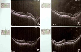
Diabetic macular oedema (DME) is the most frequent cause of visual loss among diabetic patients. The accumulation of comorbidities makes the ageing diabetic more fragile, especially in oldest-old adults, and is linked to increased hospitalisation and death rates. This case report analyses the complex treatment of Proliferative Diabetic Retinopathy with focal Photocoagulation of DME in an oldest-old woman who initially arrived with numerous diabetic comorbidities. An 82-year-old woman visited ophthalmology OPD twelve years before to perform cataract surgery. During that time, the patient did not have diabetic retinopathy at the baseline. One year before, due to irregular medication, she developed DME and best-corrected visual acuity (BCVA) was 6/36 in the right eye and prescribed to be under insulin therapy. Optical coherence tomography confirmed the presence of macular oedema and showed areas of retinal capillary non-perfusion and retinal leakage from numerous microaneurysms in the macular area. Following that, unilateral focal photocoagulation was planned. Despite grid laser, the macular oedema persisted and after two sessions of laser therapy, the macular oedema was reduced and the patient was satisfied with her vision. There is a need for further research on focal photocoagulation of DME among oldest-old adults.
References:
[1].
Raka, M., Daigavhane, S., Patil, A., 2022, A
clinical case report of diabetic retinopathy. Asian J Biomed Pharmaceut Sci,
12(90), 128, https://www.alliedacademies.org/articles/a-clinical-case-report-of-diabetic-retinopathy.pdf.
[2].
International Diabetes Federation,
2019, IDF Diabetes Atlas. 9th ed. Brussels, Belgium, International Diabetes
Federation, https://d-net.idf.org/en/library/542-idf-diabetes-atlas-2019-9th-edition.html.
[3].
Bhuvaneswari, R., Diviya, M.,
Subramanian, M., Maranan, R., Josphineleela, R., 2023, Hybrid generative model
for grading the severity of diabetic retinopathy images. Computer
Methods in Biomechanics and Biomedical Engineering: Imaging &
Visualization, 11(7), https://doi.org/10.1080/21681163.2023.2266048.
[4].
Farheen
Naz., JenilaRani D., 2022, Detection and Comparison of Diabetic Retinopathy
using Thresholding Algorithm and CMeans Clustering Algorithm, Journal of
Pharmaceutical Negative Results. 14(4), https://www.pnrjournal.com/index.php/home/article/view/899.
[5]. Suganya, M.,
KalaBarathi, S., 2024, Association of body mass index with dietary intake and
self-care health activities of mothers after postpartum: A mixed-method
research in Chennai City, India. Int J Nutr Pharmacol Neurol Dis 14:52-7,
https://doi.org/10.4103/ijnpnd.ijnpnd_71_23.
[6].
Sridhar, S.,
Pradeep Kandhasamy, J., Sinthuja, M., Sterlin Minish, T. N., 2021, WITHDRAWN:
Diabetic retinopathy detection using convolutional nueral networks algorithm, Materials
Today: Proceedings, ISSN 2214-7853, https://doi.org/10.1016/j.matpr.2021.01.059.
[7]. Obeid
A., Su D., Patel S. N., Uhr J. H., Borkar D., Gao X., et al. 2019, Outcomes of
eyes lost to follow-up with proliferative diabetic retinopathy that received
panretinal photocoagulation versus intravitreal anti-vascular endothelial
growth factor. Ophthalmology. 126(3):407–413, https://pubmed.ncbi.nlm.nih.gov/30077614/
[8]. Angela, J., Michael
Javaheri., Hamid Hosseini., Pradeep S Prasad., 2023, Purtscher‑like retinopathy
in a 19‑year‑old with maturity‑onset diabetes of the young: a case report. Journal
of Medical Case Reports, 17:255;1-4, https://doi.org/10.1186/s13256-023-03985-z
[9].
Ahmad, S. S.,
Ghani, S. A., 2012, Florid diabetic retinopathy in a young patient. J
Ophthalmic Vis Res. 7(1):84-7, https://pmc.ncbi.nlm.nih.gov/articles/PMC3381113/
[10]. Ebrahimi, M. H.,
Gharibi, H., 2016, A case study of a patient with diabetic retinopathy. Diab
Met Syndr: Clin Res Rev, http://dx.doi.org/10.1016/j.dsx.2016.01.022
[11]. Tao Y., Jiang P., Zhao Y., Song L., Ma Y., Li Y., Wang
H., 2021, Retrospective study of aflibercept in combination therapy for
high-risk proliferative diabetic retinopathy and diabetic maculopathy. Int
Ophthalmol. 41(6):2157-2165, http://doi/10.1007/s10792-021-01773-6
[12]. Gawęcki, M., 2021, Subthreshold Diode Micropulse Laser
Combined with Intravitreal Therapy for Macular Edema-A Systematized Review and
Critical Approach. J Clin Med. 31;10(7):1394, http://doi/10.3390/jcm10071394.
[13]. Thool, A. R., Dhande, N. K., Daigavane, S. V., 2021,
Study of Correlation between Renal Function Test and Severity of Diabetic
Retinopathy in Patients with Type 2 Diabetes Mellitus, J Evolution Med
Dental Sci. 17;10(20):1511http://link.gale.com/apps/doc/A663048635/HRCA
?u=anon~81ff4892&sid=googleScholar&xid=2139b05c.
[14]. Ramakrishnan, A., Veeramani, P. A., et al, 2024, Association of Various Optical Coherence Tomographic Patterns of Diabetic Macular Edema with Central Subfield Thickness and Visual Acuity: A Cross-Sectional Observational Study, Cureus. 19,16(9), https://assets.cureus.com/uploads/original_article/pdf/294904/20241019-1529365-17tn37.pdf.
