
Control of Multidrug-Resistant Hospitalized Pathogenic Bacteria Using the Secondary Metabolites of Calotropis procera and In-silico Analysis of Bacterial Virulent Proteins
Abstract:
This study explores the multidrug-resistant pattern of
hospitalized pathogens and their pharmacological impact against the secondary
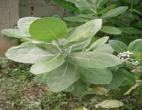
metabolites isolated from Calotropis procera, for its medicinal properties. Moreover,
this study implies that comprehensive analysis, including isolation of
multidrug-resistant hospitalized bacterial species and extraction and
characterization of secondary metabolites by GC-MS from Calotropis procera, molecular
docking, ADMET profiling, and, was conducted to evaluate the therapeutic
potential of these compounds. The multidrug-resistant Klebsiella pneumonia,
Salmonella typhi, and Pseudomonas aeruginosa were isolated and they also showed
sensitivity against C. procera leaf extract. GC-MS reveals the key volatile
compounds, including oleic acid, and the molecular docking with the proteasome
(PDB ID: 5JXG) identified 5-Methyl-2-phenylindolizine as the most promising
compound due to its high binding affinity (-6.7 kcal/mol), with
2,6-Dimethylphenol, 3,5-Dimethylaniline, and Ethyl Heptanoate showing progressively
lower affinities. Interaction analysis highlighted the importance of PRO266,
TRP531, GLU271, and ARG490 residues. ADMET profiling revealed that 2,6-dimethylphenol
and 3,5-Dimethylaniline have favorable absorption and minimal CYP interactions,
while 5-methyl-2-phenylindolizine demonstrated excellent absorption and BBB
permeability. Additionally, the study found that C. procera metabolites could
target furin, a proprotein convertase involved in bacterial virulence, offering
a novel approach to combat multidrug-resistant bacterial infections. These
findings underscore the potential of Calotropis procera metabolites as
effective therapeutic agents and active against multidrug-resistant bacterial
species.
References:
[1]. Salam MA, Al-Amin MY, Salam MT, Pawar JS, Akhter N, Rabaan AA, et al.
2023., Antimicrobial Resistance: A Growing Serious Threat for Global Public
Health. Healthcare.11: 1946. doi:10.3390/healthcare11131946
[2]. Wang J, Xu S, Zhao K, Song G, Zhao S, Liu R., 2023, Risk control of
antibiotics, antibiotic resistance genes (ARGs) and antibiotic-resistant
bacteria (ARB) during sewage sludge treatment and disposal: A review. Sci Total
Environ. 877: 162772. doi:10.1016/j.scitotenv.2023.162772
[3]. Sharma S, Chauhan A, Ranjan A, Mathkor DM, Haque S, Ramniwas S, et al.,
2024, Emerging challenges in antimicrobial resistance: implications for
pathogenic microorganisms, novel antibiotics, and their impact on
sustainability. Front Microbiol.15. doi:10.3389/fmicb.2024.1403168
[4]. Ahmed SK, Hussein S, Qurbani K, Ibrahim RH, Fareeq A, Mahmood KA, et
al., 2024, Antimicrobial resistance: Impacts, challenges, and prospects. J Med
Surgery, Public Heal. 2: 100081. doi:10.1016/j.glmedi.2024.100081
[5]. Angelini P., 2024, Plant-Derived Antimicrobials and Their Crucial Role
in Combating Antimicrobial Resistance. Antibiotics.;13: 746.
doi:10.3390/antibiotics13080746
[6]. Salam U, Ullah S, Tang Z-H, Elateeq AA, Khan Y, Khan J, et al., 2023,
Plant Metabolomics: An Overview of the Role of Primary and Secondary
Metabolites against Different Environmental Stress Factors. Life.;13: 706.
doi:10.3390/life13030706
[7]. Divekar PA, Narayana S, Divekar BA, Kumar R, Gadratagi BG, Ray A, et
al., 2022, Plant Secondary Metabolites as Defense Tools against Herbivores for
Sustainable Crop Protection. Int J Mol Sci. 23: 2690. doi:10.3390/ijms23052690
[8]. Sorescu A-A, Nuta A, Ion R-M, Iancu L., 2018, Qualitative Analysis of
Phytochemicals from Sea Buckthorn and Gooseberry. Phytochemicals - Source of
Antioxidants and Role in Disease Prevention. InTech;
doi:10.5772/intechopen.77365
[9]. Ku Y-S, Contador CA, Ng M-S, Yu J, Chung G, Lam H-M., 2020, The Effects
of Domestication on Secondary Metabolite Composition in Legumes. Front
Genet.11. doi:10.3389/fgene.2020.581357
[10]. Abeed AHA, Ali M, Ali EF., 2021, Majrashi A, Eissa MA. Induction of
Catharanthus roseus Secondary Metabolites When Calotropis procera Was Used as
Bio-Stimulant. Plants.;10: 1623. doi:10.3390/plants10081623
[11]. Wadhwani BD, Mali D, Vyas P, Nair R, Khandelwal P., 2021, A review on
phytochemical constituents and pharmacological potential of Calotropis procera.
RSC Adv. 11: 35854–35878. doi:10.1039/D1RA06703F
[12]. Habeeb A, Ramesh S, Shanmugam R., 2024, Calotropis procera and the
Pharmacological Properties of Its Aqueous Leaf Extract: A Review. Cureus.
doi:10.7759/cureus.60354
[13]. Ong CY, Ling SK, Ali RM, Chee CF, Samah ZA, Ho ASH, et al., 2009,
Systematic analysis of in vitro photo-cytotoxic activity in extracts from
terrestrial plants in Peninsula Malaysia for photodynamic therapy. J Photochem
Photobiol B Biol. 96: 216–222. doi:10.1016/j.jphotobiol.2009.06.009
[14]. Yang Y, Liang X, Jin P, Li N, Zhang Q, Yan W, et al., 2019, Screening
and determination for potential acetylcholinesterase inhibitory constituents
from ginseng stem–leaf saponins using ultrafiltration (UF)-LC-ESI-MS2.
Phytochem Anal. 30: 26–33. doi:10.1002/pca.2787
[15]. Ito A, Lai CH, Zhao X, Saito S, Hamilton MH, Appella E, et al.,
2001,p300/CBP-mediated p53 acetylation is commonly induced by p53-activating
agents and inhibited by MDM2. EMBO J. 20: 1331–1340.
doi:10.1093/emboj/20.6.1331
[16]. Rahiman FA, Mahmad N, Taha RM, Elias H, Zaman FH., 2013, Antimicrobial
properties of Lawsonia inermis syn. Lawsonia alba in vivo and in vitro. J Food,
Agric Environ. 11: 502–504.
[17]. Padmini R, Uma Maheshwari V, Saravanan P, Woo Lee K, Razia M, Alwahibi
MS, et al., 2020, Identification of novel bioactive molecules from garlic
bulbs: A special effort to determine the anticancer potential against lung
cancer with targeted drugs. Saudi J Biol Sci. 27: 3274–3289.
doi:10.1016/j.sjbs.2020.09.041
[18]. Kirubhanand C, Merciline Leonora J, Anitha S, Sangeetha R, Nachammai KT,
Langeswaran K, et al., 2023, Targeting potential receptor molecules in
non-small cell lung cancer (NSCLC) using in silico approaches. Front Mol
Biosci. 10: 1–16. doi:10.3389/fmolb.2023.1124563
[19]. Braun E, Sauter D., 2019, Furin‐mediated protein processing in
infectious diseases and cancer. Clin Transl Immunol. 8. doi:10.1002/cti2.1073
[20]. Douglas LEJ, Reihill JA, Montgomery BM, Martin SL., 2023, Furin as a
therapeutic target in cystic fibrosis airways disease. Eur Respir Rev. 32:
220256. doi:10.1183/16000617.0256-2022
[21]. Krueger E, Brown AC., 2019, Inhibition of bacterial toxin recognition of
membrane components as an anti-virulence strategy. J Biol Eng. 13: 4.
doi:10.1186/s13036-018-0138-z
[22]. do Vale A, Cabanes D, Sousa S., 2016, Bacterial Toxins as Pathogen
Weapons Against Phagocytes. Front Microbiol. 7. doi:10.3389/fmicb.2016.00042
[23]. Zhang H, Zhang Z, Li J, Qin G., 2023, New Strategies for Biocontrol of
Bacterial Toxins and Virulence: Focusing on Quorum-Sensing Interference and
Biofilm Inhibition. Toxins (Basel). 15: 570. doi:10.3390/toxins15090570
[24]. Devi KP, Pourkarim MR, Thijssen M, Sureda A, Khayatkashani M, Cismaru
CA, et al., 2022, A perspective on the
applications of furin inhibitors for the treatment of SARS-CoV-2. Pharmacol
Reports. 74: 425–430. doi:10.1007/s43440-021-00344-x
[25]. Jaganathan R, Kumaradhas P., 2024, Structural insights into Furin enzyme
inhibition to block SARS-CoV-2 spike protein cleavage: an in-silico approach. 3
Biotech. 14: 213. doi:10.1007/s13205-024-04054-y
[26]. Morris GM, Huey R, Lindstrom W, Sanner MF, Belew RK, Goodsell DS, et
al., 2010, AutoDock4 and AutoDockTools4: Automated Docking with Selective
Receptor Flexibility. J Comput Chem. 30: 2785–2791.
doi:10.1002/jcc.21256.AutoDock4
[27]. Berman HM, Battistuz T, Bhat TN, Bluhm WF, Bourne PE, Burkhardt K, et
al., 2002, The protein data bank. Acta Crystallogr Sect D Biol Crystallogr. 58:
899–907. doi:10.1107/S0907444902003451
[28]. Kim S, Chen J, Cheng T, Gindulyte A, He J, He S, et al. 2021, PubChem in
2021: New data content and improved web interfaces. Nucleic Acids Res. 49, D1388–D1395. doi:10.1093/nar/gkaa971
[29]. Dahms SO, Arciniega M, Steinmetzer T, Huber R, Than ME., 2016, Structure
of the unliganded form of the proprotein convertase furin suggests activation
by a substrate-induced mechanism. Proc Natl Acad Sci U S A. 113: 11196–11201.
doi:10.1073/pnas.1613630113
[30]. DeLano WL., 2002, Pymol: An open-source molecular graphics tool. CCP4
Newsl Protein Crystallogr. 40: 82–92.
[31]. BIOVIA. 2016. Discovery Studio Modeling Environment. San Diego: Dassault
Systems.
[32]. Pires DE V., Blundell TL, Ascher DB., 2015, pkCSM: Predicting
Small-Molecule Pharmacokinetic and Toxicity Properties Using Graph-Based
Signatures. J Med Chem. 58: 4066–4072. doi:10.1021/acs.jmedchem.5b00104
[33]. Fiehn O., 2016, Metabolomics by Gas Chromatography–Mass Spectrometry:
Combined Targeted and Untargeted Profiling. Curr Protoc Mol Biol. 114.
doi:10.1002/0471142727.mb3004s114
[34]. Jeyasundari J, Praba P, Jacob Y, Rajendran S, Kaleeswari K., 2016, Green
Synthesis and Characterization of Silver Nanoparticles Using Mimusops elengi
Flower Extract and Its Synergistic Antimicrobial Potential. Am Chem Sci J. 12:
1–11. doi:10.9734/ACSJ/2016/23161
[35]. Deshmukh SP, Patil SM, Mullani SB, Delekar SD., 2019, Silver
nanoparticles as an effective disinfectant: A review. Mater Sci Eng C. 97:
954–965. doi:10.1016/j.msec.2018.12.102
