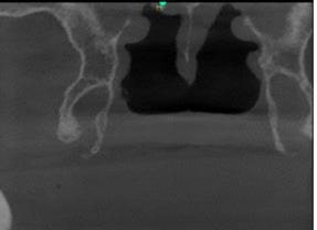
Evaluation of Pterygoid Hamulus Dimensions in Completely Edentulous Patients Using Cone Beam Computed Tomography

Abstract:
The inferior border of the medial pterygoid plate extends to form the
pterygoid hamulus (PH). The PH's length and location are crucial for these
functions. The PH’s morphology helps in interpreting the imaging and also provides
information regarding anatomical determinants to limit the posterolateral
borders of maxillary complete dentures. This can also aid in gender
identification in forensic situations. This study analyzed 80 CBCT scans from
40 male and 40 female patients (ages 25–67, median 38). Significant differences
were found in pterygoid hamulus length between sexes: males had longer hamuli
on both the right (8.840±0.299 mm vs. 7.940±0.349 mm, P=0.000) and left sides
(7.899±0.419 mm vs. 7.277±0.271 mm, P=0.002). However, no significant
differences in hamulus width were observed between males and females. These
findings suggest length variations could be useful in clinical and
anthropological contexts. Dimensions of pterygoid hamulus in completely
edentulous patients will aid the clinician in precisely recording the posterolateral
borders of maxillary dentures; this can also aid in gender determination in
fragmented skulls in forensic applications.
References:
[1].
Putz,
R., & Kroyer, A., 1999, Functional morphology of the pterygoid hamulus. Annals of Anatomy, 181(1), 85–88.
[2].
Orhan,
K., Sakul, B. U., Oz, U., & Bilecenoglu, B., 2011, Evaluation of the
pterygoid hamulus morphology using cone beam computed tomography. Oral Surgery, Oral Medicine, Oral Pathology,
Oral Radiology, and Endodontics, 112(2), e48–55.
[3].
Motiwala,
I. A., & Bathina, T., 2022, A radiographic study on pterygoid implants with
hamulus as a landmark for engaging the pterygoid plate - A retrospective study.
Annals of Maxillofacial Surgery,
12(2), 190.
[4].
Ahmed
Khan, H. L., Murthykumar, K., Sekaran, S., & Ganapathy, D., 2023, Digital
panoramic radiographs for age prediction utilizing the Tooth Coronal Index of
first mandibular bicuspids among the south Indian population. Cureus, 15(9), e45870.
[5].
Devi,
V. A., Sivakumar, N., & Ganapathy, D., 2021, Location of greater palatine
foramen in dry human skulls. International
Journal of Dentistry and Oral Science, 8(1), 1419–1421.
[6].
Kumar,
A. S., Ganapathy, D., & Duraisamy, R., 2022, Awareness on mastoid implants
among dental students. Journal of
Pharmacy & Negative Results, 13(10), 461–466.
[7].
Kende, P., Aggarwal, N., Meshram, V., Landge, J., Nimma, V.,
Mathai, P., 2019, The pterygoid hamulus syndrome–An important differential in
orofacial pain. Contemporary Clinical
Dentistry, 1;10(3), 571-6.
[8].
Oz,
U., Orhan, K., Aksoy, S., Ciftci, F., Özdoğanoğlu, T., & Rasmussen, F.,
2016, Association between pterygoid hamulus length and apnea hypopnea index in
patients with obstructive sleep apnea: A combined three-dimensional cone beam
computed tomography and polysomnographic study. Oral Surgery, Oral Medicine, Oral Pathology, Oral Radiology,
121(3), 330–339.
[9].
Bindhoo,
Y. A., Thirumurthy, V. R., Jacob, S. J., Anjanakurien, F. N. U., & Limson,
K. S., 2011, Posterior palatal seal: A literature review. International Journal of Prosthodontics and Restorative Dentistry,
1(2), 108–114.
[10]. Silverman, S. I., 1971, Dimensions and displacement patterns
of the posterior palatal seal. Journal of
Prosthetic Dentistry, 25(5), 470–488.
[11]. Winland, R. D., & Young, J. M., 1973, Maxillary complete
denture posterior palatal seal: Variations in size, shape, and location. Journal of Prosthetic Dentistry, 29(3),
256–261.
[12]. Mehra, A., Karjodkar, F. R., Sansare, K., Kapoor, R.,
Tambawala, S., & Saxena, V. S., 2021, Assessment of the dimensions of the
pterygoid hamulus for establishing age- and sex-specific reference standards
using cone-beam computed tomography. Imaging
Science in Dentistry, 51(1), 49–54.
[13]. Romoozi, E., Razavi, S. H., Barouti, P., & Rahimi, M.,
2018, Investigating the morphologic indices of the hamulus pterygoid process
using the CBCT technique. Journal of
Research in Medical and Dental Science, 6(2), 240–244.
[14]. Kachhara, S., Nallaswamy, D., Ganapathy, D., & Ariga,
P., 2021, Comparison of the CBCT, CT, 3D printing, and CAD-CAM milling options
for the most accurate root form duplication required for the root analogue
implant (RAI) protocol. Journal of the
Indian Academy of Oral Medicine and Radiology, 33(2), 141–145.
[15]. Prakash, M. S., Ganapathy, D. M., & Nesappan, T., 2019,
Assessment of labial alveolar bone thickness in maxillary central incisor and
canine in Indian population using cone-beam computed tomography. Drug Invention Today, 11(3).
[16]. Mahipathy, S. R., Jesudasan, J. S., V. S. A. C., Durairaj,
A. R., & Ananthappan, M., 2023, Misplaced pterygoid implant removed
following surgical exploration. Journal
of Evolution of Medical and Dental Sciences, 12(24), 205–207.
[17]. Shivanni, S. S., & Babu, K. Y., 2016, An anatomical
study of occurrence of pterygospinous bar in Indian skulls. Research Journal of Pharmacy and Technology,
9(8), 1166–1168.
[18]. Navaneethan, A., & Varghese, R., 2021, Comparison
between antegonial notch depth, symphysis morphology, and ramus morphology
among different growth patterns in skeletal Class I and Class II subjects. International Journal of Dentistry and Oral
Science, 8(1), 1510–1517.
[19]. Chithralekha, B., Duraisamy, R.,
Ganapathy, D. M., Maiti, S., 2022, Awareness on Pterygoid Implant Among Dental
Undergraduates. Journal for Educators,
Teachers and Trainers, 13(6). 423-430. DOI: [10.47750/jett.2022.13.06.040]
[20]. Orhan, K., Sakul, B. U. Oz. U.,
Bilecenoglu, B.,2011, Evaluation of the pterygoid hamulus morphology using cone
beam computed tomography. Oral Surgery,
Oral Medicine, Oral Pathology, Oral Radiology, and Endodontology. 1;112(2),
e48-55

