Biogenic Selenium Nanoparticles Loaded Alginate-Gelatin Scaffolds for Potential Tissue Engineering Applications

Abstract:
Selenium nanoparticles (SeNPs) were reported
for its anticancer and antimicrobial properties. Alginate and gelatin scaffolds
can act as an important biomaterial, more specifically in bone tissue engineering.
Green synthesis of SeNPs from Luffa cylindrica (LC) and loading of SeNPS with alginate-gelatin
scaffold and to check its biocompatibility. The SeNPs were prepared via the green
synthesis method and loaded into an alginate-gelatin scaffold. Characterization
studies such as UV-Vis spectroscopy, FTIR, and SEM were carried out in LC-SeNPs
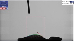
and Se-NPs loaded scaffold. The hydrophilicity of the scaffolds was determined using
water contact angle measurements. Annexin V PI assay was conducted to determine
the biocompatible nature of prepared SeNPs-loaded alginate-gelatin scaffolds. The
UV-VIS spectrum gave an intense peak at 266 and 384 nm, whereas the FTIR gave a
strong peak at 3500-500 cm-1 fingerprint regions. SEM images showed flower-shaped
LC-SeNPs and their distribution of SeNPs on the surface of alginate-gelatin scaffolds.
water contact angle measurement was found to be 29.21°. Cell viability results showed
78.11% viable cells following treatment with alg-gel-Se scaffold, revealing its
biocompatibility towards peripheral blood mononuclear cells. Overall, it could be
concluded that the SeNP-loaded alg-gel scaffold is a promising candidate for tissue
engineering, but further studies are required to confirm its potential role.
References:
[1]
Johnson, J., Shanmugam, R., and Lakshmi, T. 2022. A review on plant-mediated selenium nanoparticles
and its applications. Journal of population
therapeutics and clinical pharmacology, 28(2): e29–e40.
[2] Ingole, A. R., Thakare, S. R., Khati, N. T., Wankhade,
A. V., and Burghate, D. K. 2010. Green synthesis of selenium nanoparticles under
ambient condition. Chalcogenide Lett,
7(7): 485–489.
[3]
Naaziya, M., Biju, T. S., Francis, A. P., Veeraraghavan, V. P., Gayathri,
R., and Sankaran,
K. (2023). Synthesis, Characterization and in-vitro
Biological Studies of Curcumin decorated Biogenic Selenium Nanoparticles. Nano LIFE. https://doi.org/10.1142/s1793984423500137
[4]
Zhao, L., Wang, H., and Du, X. 2021. The therapeutic use of quercetin in ophthalmology:
recent applications. Biomedicine & Pharmacotherapy.
https://doi.org/10.1016/j.biopha.2021.111371
[5] Khurana, A., Tekula, S., Saifi, M. A., Venkatesh,
P., and Godugu,
C. 2019. Therapeutic applications of selenium nanoparticles. Biomedicine & pharmacotherapy 111: 802–812.
[6]
Jayavarsha, V., Rajeshkumar, S., Lakshmi, T., and Sulochana, G. 2022. Green synthesis
of selenium nanoparticles study using clove and cumin and its anti- inflammatory
activity. Journal of complementary medicine
research, 13(5): 84.
[7]
Skalickova, S., Milosavljevic, V., Cihalova, K., Horky, P., Richtera,
L., and Adam, V.
2017. Selenium nanoparticles as a nutritional supplement. Nutrition , 33: 83–90.
[8]
Sneka, and Santhakumar, P. 2021. Antibacterial Activity of Selenium Nanoparticles
extracted from Capparis decidua against Escherichia coli and Lactobacillus Species.
Journal of advanced pharmaceutical technology
& research, 14(8): 4452–4454.
[9]
Ndwandwe, B. K., Malinga, S. P., and Kayitesi, E. 2021. Advances in
green synthesis of selenium nanoparticles and their application in food packaging
International Journal of Food Science &
Technology https://doi.org/10.1111/ijfs.14916
[10]
Ali, S. J., Preetha, Jeevitha, Prathap, L., and Rajeshkumar. 2020. Antifungal Activity
of Selenium Nanoparticles Extracted from Capparis decidua Fruit against Candida
albicans. Journal of evolution of medical
and dental sciences, 9(34): 2452–2455. https://doi.org/10.14260/jemds/2020/533
[11]
Partap, S., Kumar, A., Sharma, N. K., and Jha, K. K. 2012. Luffa Cylindrica
: An important medicinal plant. J.
Nat. Prod. Plant Resour., 2(1):127-134
[12] Al-Snafi, A. E. 2019. Constituents and pharmacology
of Luffa cylindrica-A review. IOSR Journal
of Pharmacy. 9(9):
68-79
[13] Akinwumi, K. A., Eleyowo, O. O., and Oladipo, O.O. 2021. A review on
the ethnobotanical uses, phytochemistry and pharmacology effects of Luffa cylindrica. Natural
Drugs from Plants. https://doi.org/10.5772/intechopen.98405
[14]
Aboh, M. I., Okhale, S. E., & K., I. (2012). Preliminary studies
on Luffa cylindrica: Comparative phytochemical and antimicrobial screening of the
fresh and dried aerial parts. African journal
of microbiology research, 3088, 3091.
https://doi.org/10.5897/AJMR11.301
[15]
Balakrishnan, N., and
Sharma, A. 2013.
Preliminary phytochemical and pharmacological activities of Luffa cylindrica L.
fruit. Asian journal of pharmaceutical and
clinical research, 6(2): 113–116.
[16]
Soni, M., Gayathri, R., Sankaran, K., Veeraraghavan, V. P., and Francis, A. P. 2023. Green synthesis
of selenium nanoparticles using luffa cylindrica
and its biocompatibility for potential biomedical applications. Nano. 18(6): 2350042
[17]
Cuadros, T. R., Erices, A. A., and Aguilera, J. M. 2015. Porous matrix
of calcium alginate/gelatin with enhanced properties as scaffold for cell culture.
Journal of the mechanical behavior of biomedical
materials, 46: 331–342.
[18]
Wang, L., Zhang, H. J., Liu, X., Liu, Y., Zhu, X., Liu, X., and You, X. 2021. A Physically Cross-Linked
Sodium Alginate–Gelatin Hydrogel with High Mechanical Strength. ACS Applied Polymer Materials, 3(6): 3197–3205.
[19] You, F., Wu, X., and Chen, X. 2017. 3D printing of porous
alginate/gelatin hydrogel scaffolds and their mechanical property characterization.
International Journal of Polymeric Materials
and Polymeric Biomaterials, 66(6): 299–306.
[20]
Serafin, A., Murphy, C., Rubio, M. C., and Collins, M. N. 2021. Printable
alginate/gelatin hydrogel reinforced with carbon nanofibers as electrically conductive
scaffolds for tissue engineering. Materials
science & engineering. C, Materials for biological applications, 122: 111927.
[21] Afjoul, H., Shamloo, A., and Kamali, A. 2020. Freeze-gelled
alginate/gelatin scaffolds for wound healing applications: An in vitro, in vivo
study. Materials science & engineering.
C, Materials for biological applications, 113, 110957.
[22]
Luo, Y., Li, Y., Qin, X., Wa, Q. 2018. 3D printing of concentrated
alginate/gelatin scaffolds with homogeneous nano apatite coating for bone tissue
engineering. Materials & design, 146: 12–19.
[23] Bose, S., Roy, M., and Bandyopadhyay, A. 2012. Recent
advances in bone tissue engineering scaffolds. Trends in biotechnology, 30(10):
546–554.
[24] Gour, A., and Jain, N. K. 2019. Advances in green synthesis of nanoparticles.
Artificial cells, nanomedicine, and biotechnology
, 47(1): 844–851.
[25]
Hussain, I., Singh, N. B., Singh, A., Singh, H., and Singh, S. C. 2016. Green synthesis
of nanoparticles and its potential application. Biotechnology letters, 38(4),
545–560.
[26] Alagesan, V., and Venugopal, S. 2019. Green Synthesis
of Selenium Nanoparticle Using Leaves Extract of Withania somnifera and Its Biological
Applications and Photocatalytic Activities. BioNanoScience,
9(1):
105–116.
[27]
Sowndarya, P., Ramkumar, G., and Shivakumar, M. S. 2017. Green synthesis
of selenium nanoparticles conjugated Clausena dentata plant leaf extract and their
insecticidal potential against mosquito vectors. Artificial cells, nanomedicine, and biotechnology , 45(8): 1490–1495.
[28] Fardsadegh, B., and Jafarizadeh-Malmiri, H. 2019. Aloe
vera leaf extract mediated green synthesis of selenium nanoparticles and assessment
of their in vitro antimicrobial activity against spoilage fungi and pathogenic bacteria
strains. Green Processing and Synthesis,
8(1):
399–407.
[29]
Sharma, A., Puri, V., Kumar, P., and Singh, I. 2020. Rifampicin-Loaded
Alginate-Gelatin Fibers Incorporated within Transdermal Films as a Fiber-in-
Film System for Wound Healing Applications. Membranes,
11(1):7
[30]
Leena, R. S., Vairamani, M., and Selvamurugan, N. 2017. Alginate/Gelatin
scaffolds incorporated with Silibinin-loaded Chitosan nanoparticles for bone formation
in vitro. Colloids and surfaces. B, Biointerfaces,
158, 308–318.

