
Curcumin Coated Orthosiphon Stamineus Leaf Extract Based Selenium Nanoparticle for Potential Tissue Engineering Applications
Abstract:
Curcumin is a
type of polyphenol phytochemical that is bright yellow in colour and produced by
the plant Curcuma longa. Despite
its pharmacological properties, curcumin has low bioavailability, poor solubility,
and undergoes rapid degradation. Nanoparticles (NPs) are used as a nanocarrier for
drug delivery, to improve the stability and pharmacokinetics of the drug. Therefore,
by coating curcumin over selenium NPs (SeNPs), the bioactivity, bioavailability,
stability, and may increase the solubility of curcumin of SeNPs. This study
aimed to synthesize the SeNPs from Orthosiphon stamineus leaf extract and coat it
with curcumin and to characterize it and check its biocompatibility. Biosynthesis of SeNPs was carried out using
plant extract of Evolvulus alsinoides and characterized using UV spectrophotometer,
FT-IR, and SEM. Annexin V PI apoptotic and Hemolytic assay were used for checking
biocompatibility. The UV-Vis spectrum
gave a strong peak at 265 and 423 nm at various time intervals, indicating the SeNPs
formation. Similarly, FT-IR has strong absorption bands at 3279, 1284, 1072, 1028,
and cm−1
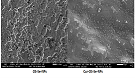
with wavelengths ranging from 4000-500 cm-1. SEM analysis of biosynthesized
SeNPs showed a spherical shape. Our results suggest that curcumin-coated SeNPs possess
greater biocompatibility towards PBMCs which was evaluated by Annexin V - PI assay
and erythrocytes by hemolytic studies. Curcumin-coated Selenium nanoparticles were
successfully synthesized by the biological method using leaf extract of Orthosiphon
stamineus and reported as biocompatible using Flow cytometry. But a more detailed
study should be done for implementing it in tissue engineering.
References:
[1]
Nabi, F., Arain, M. A., Hassan, F., Umar, M., Rajput, N., Alagawany,
M., … Liu, J. 2020. Nutraceutical role of selenium nanoparticles in poultry nutrition:
a review. Worlds Poult. Sci. J. 76 (3): 459–471.
[2]
Pandiyan, I., Sri, S. D., Indiran, M. A., Rathinavelu, P. K., Prabakar,
J., and Rajeshkumar,
S. (2022). Antioxidant, anti-inflammatory activity of Thymus vulgaris-mediated selenium
nanoparticles: An in vitro study. J. Conserv. Dent. 25 (3):
241–245.
[3]
Sushanthi, S., Srisakthi, D., MeignanaArumugham, I., Pradeepkumar,
R., and Rajeshkumar,
S. 2021. Vernonia Amygdalina Mediated Copper Nanoparticles and its Characterization
and Antimicrobial Activity - An In Vitro Study. Int J Dent. Oral Sci., 8 (7): 3330–3334.
[4]
Maiyo, F., and Singh, M. 2017. Selenium nanoparticles: potential in cancer
gene and drug delivery. Nanomedicine ,
12 (9): 1075–1089.
[5]
Maliael, M. T., Jain, R. K., and Srirengalakshmi, M. 2022. Effect
of nanoparticle coatings on frictional resistance of orthodontic archwires: a systematic
review and meta-analysis. World J. Dent,
13(4), 417–424.
[6]
Nasim, I., Jabin, Z., Kumar, S. R., and Vishnupriya, V. (2022). Green synthesis
of calcium hydroxide-coated silver nanoparticles using Andrographis paniculata and
Ocimum sanctum Linn. leaf extracts: An antimicrobial and cytotoxic activity. JJ. Conserv. Dent. 25 (4): 369–374.
[7]
Kamath, K. A., Nasim, I., and Rajeshkumar, S. (2020). Evaluation
of the re-mineralization capacity of a gold nanoparticle-based dental varnish: An
in vitro study. J. Conserv. Dent. 23 (4): 390–394.
[8]
Maheswari, T. N. U., and Dhanvanth, M. 2022. Topical herbal therapeutic formulation
used in the management of oral potentially malignant disorders – A systematic review.
J. Indian Acad. Oral Med. Radiol. 34 (2): 223.
[9]
Ghaderi, R. S., Adibian, F., Sabouri, Z., Davoodi, J., Kazemi, M.,
Amel Jamehdar, S., … Daroudi, M. 2022. Green synthesis of selenium nanoparticle
by Abelmoschus esculentus extract and assessment of its antibacterial activity.
Mater. Tech. 37(10), 1289–1297.
[10]
Deng, W., Xie, Q., Wang, H., Ma, Z., Wu, B., and Zhang,
X. 2017. Selenium nanoparticles as versatile carriers for oral delivery of insulin:
Insight into the synergic antidiabetic effect and mechanism. Nanomed.: Nanotechnol. Biol. Med. 13 (6): 1965–1974.
[11]
Pyrzynska, K., and Sentkowska, A. 2022. Biosynthesis of selenium nanoparticles
using plant extracts. J. Nanostructure
Chem.12 (4): 467–480.
[12]
Sentkowska, A., and Pyrzyńska, K. 2022. The Influence of Synthesis Conditions on
the Antioxidant Activity of Selenium Nanoparticles. Molecules , 27 (8): 2486
[13]
Shirmehenji, R., Javanshir, S., and Honarmand, M. 2021. A Green Approach
to the Bio-based Synthesis of Selenium Nanoparticles from Mining Waste. J. Cluster Sci. 32 (5): 1311–1323.
[14]
Kora, A. J., and Rastogi, L. 2016. Biomimetic synthesis of selenium nanoparticles
by Pseudomonas aeruginosa ATCC 27853: An approach for conversion of selenite. J. Environ. Manage. 181: 231–236.
[15]
Meenambigai, K., Kokila, R., Chandhirasekar, K., Thendralmanikandan,
A., Kaliannan, D., Ibrahim, K. S., … Nareshkumar, A. 2022. Green Synthesis of Selenium
Nanoparticles Mediated by Nilgirianthus ciliates
Leaf Extracts for Antimicrobial Activity on Foodborne Pathogenic Microbes and Pesticidal
Activity Against Aedes aegypti with Molecular Docking. Biol.
Trace Elem. Res.
200 (6): 2948–2962.
[16]
Hatami, R., Javadi, A., & Jafarizadeh-Malmiri, H. (2020). Effectiveness
of six different methods in green synthesis of selenium nanoparticles using propolis
extract: Screening and characterization. Green
Processing and Synthesis, 9(1), 685–692.
[17]
Johnson, J., Shanmugam, R., & Lakshmi, T. (2022). A review on plant-mediated
selenium nanoparticles and its applications. J. Popul. Ther. Clin. Pharmacol. 28 (2): e29–e40.
[18]
Mi, X.-J., Choi, H. S., Perumalsamy, H., Shanmugam, R., Thangavelu,
L., Balusamy, S. R., and Kim, Y.-J. 2022. Biosynthesis and cytotoxic effect of silymarin-functionalized
selenium nanoparticles induced autophagy mediated cellular apoptosis via downregulation
of PI3K/Akt/mTOR pathway in gastric cancer. Phytomedicine,
99: 154014.
[19]
Ezhilarasan, D., Lakshmi, T., and Mallineni, S. K. 2022. Nano-based
targeted drug delivery for lung cancer: therapeutic avenues and challenges. Nanomedicine , 17 (24): 1855–1869.
[20]
Safaei, M., Mozaffari, H. R., Moradpoor, H., Imani, M. M., Sharifi,
R., and Golshah,
A. 2022. Optimization of Green Synthesis of Selenium Nanoparticles and Evaluation
of Their Antifungal Activity against Oral Candida
albicans Infection. Adv. Mater.
Sci. Eng. 2022: 1376998
[21]
Liu, W., Li, X., Wong, Y.-S., Zheng, W., Zhang, Y., Cao, W., and Chen, T.
2012. Selenium nanoparticles as a carrier of 5-fluorouracil to achieve anticancer
synergism. ACS nano, 6 (8): 6578–6591.
[22]
Nasim, I., Rajesh Kumar, S., Vishnupriya, V.,
and Jabin, Z. 2020. Cytotoxicity and anti-microbial analysis of silver and graphene
oxide bio nanoparticles. Bioinformation,
16 (11): 831–836.
[23]
Nasim, I., Rajeshkumar, S., and Vishnupriya,
V. 2021. Green synthesis of reduced graphene oxide nanoparticles, its characterization,
and antimicrobial properties against common oral pathogens. Int J Dentistry Oral Sci., 8 (2): 1670–1675.
[24]
Kishore, S. O. G., Priya, A. J., and Narayanan,
L. 2020. Controlling of oral pathogens using turmeric and tulsi herbal formulation
mediated copper nanoparticles. Plant Cell
Biotechnol. Mol. Biol. 21 (53-54): 33-37
[25]
Rieshy, V., Priya, J., Arivarasu, L., and Kumar,
S. R. 2020. Enhanced Antimicrobial Activity of Herbal Formulation Mediated Copper
Nanoparticles Against Clinical Pathogens. Plant
Cell Biotechnol. Mol. Biol. 21 (53-54): 52-56.
[26]
Sneka, and Santhakumar, P. 2021. Antibacterial Activity of Selenium Nanoparticles
extracted from Capparis decidua against Escherichia coli and Lactobacillus Species.
J. Adv. Pharm. Tech. Res. 14 (8): 4452–4454.
[27]
Palombo, E. A. 2011. Traditional Medicinal Plant Extracts and Natural
Products with Activity against Oral Bacteria: Potential Application in the Prevention
and Treatment of Oral Diseases. Evid. Based. Complement. Alternat. Med. 2011: 680354.
[28]
Rajeshkumar, S., Lakshmi, T., and Tharani, M. (2021). Green synthesis
of copper nanoparticles synthesized using black tea and its antibacterial activity
against oral pathogens. Int. J. Dent. Oral
Sci., 8 (9): 4156–4159.
[29]
Rajeshkumar, S., and Lakshmi, S. 2021. Anticariogenic activity of silver nanoparticles
synthesized using fresh leaves extract of kalanchoe pinnata. . Int J Dentistry Oral Sci., 8 (7): 2985–2987.
[30]
Twaij, B. M., and Hasan, M. N. 2022. Bioactive Secondary Metabolites from Plant
Sources: Types, Synthesis, and Their Therapeutic Uses. Int. J.Plant Biol. 13 (1): 4–14.
[31]
Sharifi-Rad, J., Rayess, Y. E., Rizk, A. A., Sadaka, C., Zgheib, R.,
Zam, W., … Martins, N. 2020. Turmeric and Its Major Compound Curcumin on Health:
Bioactive Effects and Safety Profiles for Food, Pharmaceutical, Biotechnological
and Medicinal Applications. Front. Pharmacol. 11: 01021.
[32]
Ashraf, K., Sultan, S., and Adam, A. 2018. Orthosiphon stamineus Benth. is an Outstanding
Food Medicine: Review of Phytochemical and Pharmacological Activities. J. Pharm. Bioallied Sci. 10 (3): 109–118.
[33]
Wang, Q., Wang, J., Li, N., Liu, J., Zhou, J., Zhuang, P., and Chen, H.
2022. A Systematic Review of Orthosiphon stamineus Benth. in the Treatment of Diabetes
and Its Complications. Molecules. 27 (2):
444
[34]
Hossain, M. A., Ismail, Z., Rahman, A., and Kang, S. C. 2008. Chemical composition
and anti-fungal properties of the essential oils and crude extracts of Orthosiphon
stamineus Benth. Ind. Crops Prod. 27 (3): 328–334.
[35]
Hossain, M. A., and Mizanur Rahman, S. M. 2015. Isolation and characterisation
of flavonoids from the leaves of medicinal plant Orthosiphon stamineus. Arab. J. Chem. 8 (2): 218–221.
[36]
Ameer, O. Z., Salman, I. M., Asmawi, M. Z., Ibraheem, Z. O., and Yam, M.
F. 2012. Orthosiphon stamineus: traditional uses, phytochemistry, pharmacology,
and toxicology. J. Med. Food. 15 (8):, 678–690.
[37]
Guo, M., Li, Y., Lin, Z., Zhao, M., Xiao, M., Wang, C., … Zhu, B. 2017.
Surface decoration of selenium nanoparticles with curcumin induced HepG2 cell apoptosis
through ROS mediated p53 and AKT signaling pathways. RSC Adv. 7 (83): 52456–52464.
[38]
Francis, S., Joseph, S., Koshy, E. P., and Mathew, B. 2017. Green synthesis
and characterization of gold and silver nanoparticles using Mussaenda glabrata leaf
extract and their environmental applications to dye degradation. Environ.
Sci. Pollut. Res. 24 (21): 17347–17357.
[39]
Bulmus, V., Woodward, M., Lin, L., Murthy, N., Stayton, P., and Hoffman,
A. 2003. A new pH-responsive and glutathione-reactive, endosomal membrane-disruptive
polymeric carrier for intracellular delivery of biomolecular drugs. J. Control. Release. 93(2), 105–120.
[40]
Qi, B., Wang, C., Ding, J., and Tao, W. (2019). Editorial: Applications
of Nanobiotechnology in Pharmacology. Front. Pharmacol. 10: 1451.
[41]
Cittrarasu, V., Kaliannan, D., Dharman, K., Maluventhen, V., Easwaran,
M., Liu, W. C.,
Arumugam, M. 2021. Green synthesis of selenium nanoparticles mediated from Ceropegia
bulbosa Roxb extract and its cytotoxicity, antimicrobial, mosquitocidal and photocatalytic
activities. Sci. Rep. 11 (1): 1032.
[42]
Gunti, L., Dass, R. S., and
Kalagatur, N. K. 2019. Phytofabrication of Selenium Nanoparticles
from Emblica officinalis Fruit Extract and Exploring Its Biopotential Applications:
Antioxidant, Antimicrobial, and Biocompatibility. Front. Microbiol. 10: 931.
[43]
Ramamurthy, C. H., Sampath, K. S., Arunkumar, P., Suresh Kumar, M.,
Sujatha, V., Premkumar, K., and Thirunavukkarasu, C. 2013. Green synthesis and characterization
of selenium nanoparticles and its augmented cytotoxicity with doxorubicin on cancer
cells. Bioprocess Biosyst. Eng. 36 (8): 1131-1139.
