
Synthesis, Characterisation, and in Vitro Biocompatibility Studies of Selenium Nanoparticles Synthesized using Hybanthus Enneaspermus Plant Extract for Potential Biomedical Applications
Abstract:
Hybanthus enneaspermus (HE) is a traditional medicinal plant
used for treating various disease conditions. Selenium nanoparticles (SeNPs) possess
various properties such as anticancer, antioxidant, etc. The objective of the
present study is to conduct green synthesis of selenium nanoparticles using Hybanthus
enneaspermus(HE) and evaluate their biocompatibility. Leaves of HE are utilized
for synthesizing SeNPs. Characterization studies of HE-SeNPs are carried out using
UV spectrophotometry, FT-IR spectroscopy, and SEM. To check the biocompatibility,
hemolytic assay, and Annexin V-PI assays are carried out. A change in color is observed
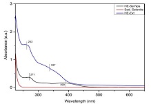
after the addition of sodium selenite to the leaf extract. UV spectrophotometry
gives a peak at 271 nm confirming the synthesis of SeNPs. FT-IR gives peaks at 3224,
1565, 1399, 1078, 784, and 717 cm-1 with a fingerprint of 3500 - 1000
cm-1. SEM analysis shows the spherical morphology of the SeNPs. HE-SeNPs
at lower concentrations cause less hemolysis. However, HE-SeNPs are found to be
less biocompatible, so further studies are needed to confirm their biocompatible
nature. SeNPs synthesized from HE can be ideal for biomedical applications but further
studies are required to check its biocompatibility.
References:
[1]
Wacker, M. G. (2014). Nanotherapeutics—Product Development Along the
“Nanomaterial” Discussion. Journal of pharmaceutical
sciences, 103(3), 777–784. https://doi.org/10.1002/jps.23879.
[2]
Abbasian, R., & Jafarizadeh-Malmiri, H. (2020). Green approach
in gold, silver and selenium nanoparticles using coffee bean extract. Open Agriculture, 5(1), 761–767. https://doi.org/10.1515/opag-2020-0074.
[3]
Fardsadegh, B., & Jafarizadeh-Malmiri, H. (2019). Aloe vera leaf
extract mediated green synthesis of selenium nanoparticles and assessment of their
In vitro antimicrobial activity against spoilage fungi and pathogenic bacteria strains.
Green Processing and Synthesis, 8(1), 399–407. https://doi.org/10.1515/gps-2019-0007.
[4]
Nasim, I., Rajeshkumar, S., & Vishnupriya, V. (2021). Green synthesis
of reduced graphene oxide nanoparticles, its characterization, and antimicrobial
properties against common oral pathogens. Int
J Dentistry Oral Sci., 8(2), 1670–1675.
[5]
Nasim, I., Rajesh Kumar, S., Vishnupriya, V., & Jabin, Z. (2020).
Cytotoxicity and anti-microbial analysis of silver and graphene oxide bio nanoparticles.
Bioinformation, 16(11), 831–836. https://doi.org/10.6026/97320630016831.
[6]
Fritea, L., Laslo, V., Cavalu, S., Costea, T., & Vicas, S. I. (2017).
Green biosynthesis of selenium nanoparticles using parsley (Petroselinum crispum)
leaves extract. Studia Universitatis” Vasile
Goldis” Arad. Seria Stiintele Vietii (Life Sciences Series), 27(3), 203–208. Retrieved from http://www.studiauniversitatis.ro/pdf/27-%202017/27-3-2017/7-%20SUVG-27-3-%20L.F.-%20203-208.pdf.
[7]
Kishore, S. O. G., Priya, A. J., & Narayanan, L. (n.d.). Controlling
of oral pathogens using turmeric and tulsi herbal formulation mediated copper nanoparticles.
Plant cell biotechnology and molecular biology.
[8]
Rieshy, V., Priya, J., Arivarasu, L., & Kumar, S. R. (n.d.). Enhanced
Antimicrobial Activity of Herbal Formulation Mediated Copper Nanoparticles
Against Clinical Pathogens. The Plant cell.
[9]
Rajeshkumar, S., & Lakshmi, S. (2021). Anticariogenic activity
of silver nanoparticles synthesized using fresh leaves extract of kalanchoe pinnata.
. Int J Dentistry Oral Sci., 8(7), 2985–2987.
[10]
Rajeshkumar, S., Lakshmi, T., & Tharani, M. (2021). Green synthesis
of copper nanoparticles synthesized using black tea and its antibacterial activity
against oral pathogens. Int. J. Dent. Oral
Sci., 8(9), 4156–4159.
[11]
Maheswari, T. N. U., & Dhanvanth, M. (2022). Topical herbal therapeutic
formulation used in the management of oral potentially malignant disorders – A systematic
review. Journal of Indian Academy of Oral
Medicine and Radiology, 34(2), 223.
https://doi.org/10.4103/jiaomr.jiaomr_101_21.
[12]
Radomska, D., Czarnomysy, R., Radomski, D., Bielawska, A., & Bielawski,
K. (2021). Selenium as a Bioactive Micronutrient in the Human Diet and Its Cancer
Chemopreventive Activity. Nutrients, 13(5). https://doi.org/10.3390/nu13051649
[13]
Souza, L. M. dos S., Dibo, M., Sarmiento, J. J. P., Seabra, A. B.,
Medeiros, L. P., Lourenço, I. M., … Nakazato, G. (2022). Biosynthesis of selenium
nanoparticles using combinations of plant extracts and their antibacterial activity.
Current Research in Green and Sustainable
Chemistry, 5, 100303. https://doi.org/10.1016/j.crgsc.2022.100303.
[14]
Johnson, J., Shanmugam, R., & Lakshmi, T. (2022). A review on plant-mediated
selenium nanoparticles and its applications. Journal of population therapeutics and clinical pharmacology = Journal de
la therapeutique des populations et de la pharamcologie clinique, 28(2), e29–e40. https://doi.org/10.47750/jptcp.2022.870.
[15]
Mi, X.-J., Choi, H. S., Perumalsamy, H., Shanmugam, R., Thangavelu,
L., Balusamy, S. R., & Kim, Y.-J. (2022). Biosynthesis and cytotoxic effect
of silymarin-functionalized selenium nanoparticles induced autophagy mediated cellular
apoptosis via downregulation of PI3K/Akt/mTOR pathway in gastric cancer. Phytomedicine: international journal of phytotherapy
and phytopharmacology, 99, 154014.
https://doi.org/10.1016/j.phymed.2022.154014.
[16]
Sneka, & Santhakumar, P. (2021). Antibacterial Activity of Selenium
Nanoparticles extracted from Capparis decidua against Escherichia coli and Lactobacillus
Species. Journal of advanced pharmaceutical
technology & research, 14(8),
4452–4454. https://doi.org/10.52711/0974-360x.2021.00773.
[17]
Pandiyan, I., Sri, S. D., Indiran, M. A., Rathinavelu, P. K., Prabakar,
J., & Rajeshkumar, S. (2022). Antioxidant, anti-inflammatory activity of Thymus
vulgaris-mediated selenium nanoparticles: An in vitro study. Journal of conservative dentistry: JCD, 25(3), 241–245. https://doi.org/10.4103/JCD.JCD_369_21.
[18]
Hosnedlova, B., Kepinska, M., Skalickova, S., Fernandez, C., Ruttkay-Nedecky,
B., Peng, Q., … Kizek, R. (2018). Nano-selenium and its nanomedicine applications:
a critical review. International journal of
nanomedicine, 13, 2107–2128. https://doi.org/10.2147/IJN.S157541.
[19]
Benstoem, C., Goetzenich, A., Kraemer, S., Borosch, S., Manzanares,
W., Hardy, G., & Stoppe, C. (2015). Selenium and its supplementation in cardiovascular
disease--what do we know? Nutrients, 7(5), 3094–3118. https://doi.org/10.3390/nu7053094.
[20]
Kamath, K. A., Nasim, I., & Rajeshkumar, S. (2020). Evaluation
of the re-mineralization capacity of a gold nanoparticle-based dental varnish: An
in vitro study. Journal of conservative dentistry:
JCD, 23(4), 390–394.
[21]
Nasim, I., Jabin, Z., Kumar, S. R., & Vishnupriya, V. (2022). Green
synthesis of calcium hydroxide-coated silver nanoparticles using Andrographis paniculata
and Ocimum sanctum Linn. leaf extracts: An antimicrobial and cytotoxic activity.
Journal of conservative dentistry: JCD,
25(4), 369–374. https://doi.org/10.4103/jcd.jcd_411_21.
[22]
Wadhwani, S. A., Gorain, M., Banerjee, P., Shedbalkar, U. U., Singh,
R., Kundu, G. C., & Chopade, B. A. (2017). Green synthesis of selenium nanoparticles
using Acinetobacter sp. SW30: optimization, characterization and its anticancer
activity in breast cancer cells. International
journal of nanomedicine, 12, 6841–6855.
https://doi.org/10.2147/IJN.S139212.
[23]
Faramarzi, S., Anzabi, Y., & Jafarizadeh-Malmiri, H. (2020). Nanobiotechnology
approach in intracellular selenium nanoparticle synthesis using Saccharomyces cerevisiae—fabrication
and characterization. Archives of microbiology,
202(5), 1203–1209. https://doi.org/10.1007/s00203-020-01831-0.
[24]
Maliael, M. T., Jain, R. K., & Srirengalakshmi, M. (2022). Effect
of nanoparticle coatings on frictional resistance of orthodontic archwires: a systematic
review and meta-analysis. World J. Dent,
13(4), 417–424. Retrieved from https://www.wjoud.com/abstractArticleContentBrowse/WJOUD/28479/JPJ/fullText.
[25]
Sushanthi, S., Srisakthi, D., MeignanaArumugham, I., Pradeepkumar,
R., & Rajeshkumar, S. (2021). Vernonia Amygdalina Mediated Copper Nanoparticles
and its Characterization and Antimicrobial Activity - An In Vitro Study. Int J Dentistry Oral Sci., 8(7), 3330–3334.
[26]
Shekhawat, M. S., & Manokari, M. (2018). In vitro multiplication,
micromorphological studies and ex vitro rooting of Hybanthus enneaspermus (L.) F.
Muell. – a rare medicinal plant. Acta Botanica
Croatica. https://doi.org/10.1515/botcro-2017-0012.
[27]
Patel, D. K., Kumar, R., Laloo, D., & Hemalatha, S. (2011). Evaluation
of phytochemical and antioxidant activities of the different fractions of Hybanthus
enneaspermus (Linn.) F. Muell. (Violaceae). Asian
Pacific journal of tropical medicine, 4(5),
391–396. https://doi.org/10.1016/S1995-7645(11)60110-7.
[28]
Patel, D. K., Kumar, R., Prasad, S. K., Sairam, K., & Hemalatha,
S. (2011). Antidiabetic and in vitro antioxidant potential of Hybanthus enneaspermus
(Linn) F. Muell in streptozotocin-induced diabetic rats. Asian Pacific journal of tropical biomedicine, 1(4), 316–322. https://doi.org/10.1016/S2221-1691(11)60051-8.
[29]
Patel, D. K., Kumar, R., Sairam, K., & Hemalatha, S. (2013). Hybanthus
enneaspermus (L.) F. Muell: a concise report on its phytopharmacological aspects.
Chinese journal of natural medicines,
11(3), 199–206. https://doi.org/10.1016/S1875-5364(13)60017-5.
[30]
Tripathy, S., Sahoo, S. P., Pradhan, D., Sahoo, S., & Satapathy,
D. K. (2009). Evaluation of anti arthritic potential of Hybanthus enneaspermus.
Retrieved February 6, 2023, from https://citeseerx.ist.psu.edu/document?repid=rep1&type=pdf&doi=b493b471da54de812289586c9fe53948b45e25f6.
[31]
Anand, T., & Gokulakrishnan, K. (2012). Phytochemical analysis
of Hybanthus enneaspermus using UV, FTIR and GC-MS. IOSR Journal of Pharmacy, 2(3),
520–524. Retrieved from https://www.researchgate.net/profile/Anand_Thirupathi2/publication/266286276_Phytochemical_analysis_of_hybanthus_enneaspermus_using_UV_FTIR_and_GC-_MS/links/55808d8808ae47061e5f3311.pdf.
[32]
Rajsekhar, P. B., Bharani, R. S. A., Angel, K. J., Ramachandran, M.,
& Rajsekhar, S. P. V. (2016). Hybanthus enneaspermus (L) F. Muell: A phytopharmacological
review on herbal medicine. Journal of chemical
and pharmaceutical research, 8(1),
351–355.
[33]
Hemalatha, S., Wahi, A. K., Singh, P. N., & Chansouria, J. P. N.
(2003). Anticonvulsant and free radical scavenging activity of Hybanthus enneaspermus:
A preliminary screening. Indian journal of
traditional knowledge. Retrieved from http://nopr.niscpr.res.in/handle/123456789/25972.
[34]
Weniger, B., Lagnika, L., Vonthron-Sénécheau, C., Adjobimey, T., Gbenou,
J., Moudachirou, M., … Sanni, A. (2004). Evaluation of ethnobotanically selected
Benin medicinal plants for their in vitro antiplasmodial activity. Journal of ethnopharmacology, 90(2-3), 279–284. https://doi.org/10.1016/j.jep.2003.10.002.
[35]
Francis, A. P., Gurudevan, S., & Jayakrishnan, A. (2018). Synthetic
polymannose as a drug carrier: synthesis, toxicity and anti-fungal activity of polymannose-amphotericin
B conjugates. Journal of biomaterials science.
Polymer edition, 29(13), 1529–1548.
https://doi.org/10.1080/09205063.2018.1469186.
[36]
Anu, K., Singaravelu, G., Murugan, K., & Benelli, G. (2017). Green-Synthesis
of Selenium Nanoparticles Using Garlic Cloves (Allium sativum): Biophysical Characterization
and Cytotoxicity on Vero Cells. Journal of
Cluster Science, 28(1), 551–563. https://doi.org/10.1007/s10876-016-1123-7.
[37]
Alagesan, V., & Venugopal, S. (2019). Green Synthesis of Selenium
Nanoparticle Using Leaves Extract of Withania somnifera and Its Biological Applications
and Photocatalytic Activities. BioNanoScience,
9(1), 105–116. https://doi.org/10.1007/s12668-018-0566-8.
