References:
[1.] Brandsma, D., Stalpers, L., Taal, W., et al (2008) .
Clinical features, mechanisms, and management of pseudoprogression in malignant
gliomas. The Lancet Oncology, 9(5), 453-461.
[2.] Brismar, J., Roberson, G.H., Davis, K.R (1976).
Radiation necrosis of the brain. Neuroradiological considerations with computed
tomography. Neuroradiology, 12(2), 109-113
[3.] Chamberlain, M.C., Glantz, M.J., Chalmers, L., et al
(2007) . Early necrosis following concurrent Temodar and radiotherapy in
patients with glioblastoma. - Journal of Neuro oncology, 82(1), 81-83.
[4.] Curnes, J., Laster, D., Bal,l M., et al (1986) . MRI
of radiation injury to the brain. Am J Roentgenol, 147(1), 119-124.
[5.] Dellen, J.v., Danzinger, A (1978). Failure of
computerized tomography to differentiate between radiation necrosis and
cerebral tumor. S Afr Med J, 53(5), 171-172.
[6.] Henson, J.W., Ulmer, S., Harris, G.J (2008). Brain
tumor imaging in clinical trials. Am J Neuroradiol, 29(3), 419-424.
[7.] Jain, R., Scarpace, L., Ellika, S., et al (2010).
Imaging response criteria for recurrent gliomas treated with bevacizumab: Role
of diffusion weighted imaging as an imaging biomarker. Journal of
Neuro-Oncology ,96(3),423-431.
[8.] Kato, T., Yutaka, S., Tada, M., et al (1996). Long
term evaluation of Radiation-induced brain damage by serial magnetic resonance
imaging. Neurologia Medico-Chirurgica, 36(12), 870-876.
[9.] Kumar, A.J., Leeds, N.E., Fuller, G.N., et al (2000).
Malignant gliomas: MR imaging spectrum of radiation therapy- and
chemotherapy-induced necrosis of the brain after treatment. Radiolog, ;217(2),
377-384.
[10.] Lyubimova, N., Hopewell , J.W, (2004). Experimental
evidence to support the hypothesis that damage to vascular endothelium plays
the primary role in the development of late radiation-induced CNS injury. Br
J Radiol; 77(918), 488-492.
[11.] Macdonald, D.R., Cascino, T.L., Schold, S.C., et al,
(1990). Response criteria for phase II studies of supratentorial malignant
glioma. Journal of Clinical Oncology, 8(7), 1277-1280.
[12.] Mikhael, M. (1978).Radiation necrosis of the brain:
correlation between computed tomography, pathology, and dose distribution. J
comput Assist Tomogr, 2(1), 71-80.
[13.] Mullins, M.E., Barest, G.D., Schaefer, P.W., et al
(2005). Radiation necrosis versus glioma recurrence: conventional MR imaging
clues to diagnosis. AJNR Am J Neuroradiol , 26(8), 1967-1972.
[14.] Sorensen, A.G., Batchelor, T.T., Wen, P.Y., et al
(2008). Response criteria for glioma. Nat Clin Prac Oncol, 5(11),
634-644.
[15.] Wen P.Y., Macdonald, D.R., Reardon, D.A., et al
(2010). Updated response assessment criteria for high-grade gliomas: response
assessment in neuro-oncology working group. Journal of Clinical Oncology,
28(11), 1963-1972.
[16.]
Zuniga,
R., Torcuator, R., Jain, R., et al (2009). Efficacy, safety and patterns of
response and recurrence in patients with recurrent high-grade gliomas treated
with bevacizumab plus irinotecan. Journal of Neuro-Oncology, 91(3),329

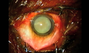 Case Report - Aniridia Lens with Manual Sutureless Medium Incision Cataract Surgery – Big Size, Big SurpriseAuthor: Jasdeep Singh Sandhu
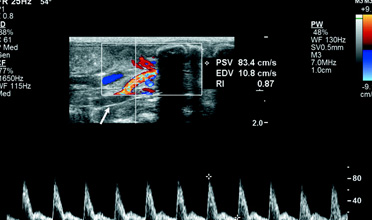 The Use of Unenhanced Doppler Sonography in the Evaluation of Solid Breast LesionsAuthor: Avineesh Skandan
 Diffusion-Weighted Imaging in the Follow-Up of Treated High-Grade Gliomas: Tumor Recurrence Versus Radiation InjuryAuthor: Vikas Rai
 Fat Embolism Syndrome in Fracture Tibia Treated by Unreamed Interlocking NailAuthor: Oral Roberts R.J.
