References:
[1] Yingchoncharoen T, Agarwal S, Popovic ZB. Normal
ranges of left ventricular strain: a meta-analysis. J Am Soc Echocardiogr.
2013; 26:185-91.
[2] Lang RM, Badano LP, Mor-Avi V, et al. Recommendations
for cardiac chamber quantification by echocardiography in adults: an update from
the American Society of Echocardiography and the European Association of Cardiovascular
Imaging. J Am Soc Echocardiogr, 2015; 28:1- 39.e14.
[3] Jasaityte R, Heyde B, D’Hooge J. Current state
of three-dimensional myocardial strain estimation using echocardiography. J Am
Soc Echocardiogr.2012; 26:15-28.
[4] Papademetris X, Sinusad AJ, Dione DP, et al.
Estimation of 3D left ventricular deformation from echocardiography. Med Image
Anal. 2001; 15:17-28.
[5] Elen A, Choi HF, Loeckx D, et al. Three-dimensional
cardiac strain estimation using spatio-temporal elastic registration of ultrasound
images: a feasibility study. IEEE Trans Med Imaging. 2008; 27:1580-91.
[6] Lower R. Tractatus de Corde, London, UK: Oxford
University Press, 1669.
[7] Kaku K, Takeuchi M, Tsang W, Takigiku K, Yasukochi
S, Patel AR, Mor Avi V, Lang RM, Otsuji Y. Age-related normal range of left ventricular
strain and torsion using three-dimensional speckle-tracking echocardiography. J
Am Soc Echocardiogr. 2014; 27:55-64. doi: 10.1016/j. echo.2013.10.002.
[8] Takahashi K, Al Naami G. Thompson R, Inage A,
Mackie AS, Smallhorn JF. Normal rotational, torsion and untwisting data in children,
adolescents, and young adults. J Am Soc Echocardiogr. 2010; 23:286-293. doi:
10.1016/j.echo.2009.11.018.
[9] A. D’ Andrea, A. Stanziola, E. Di palma, M.
Martino, M. D’ Alto, S. Dellegrottaglie. Right Ventricular structure and function
in idiopathic pulmonary fibrosis with or without pulmonary hypertension Echocardiography
33 (1) (Jun 11, 2015) 57-65.
[10] Georgette E. Hoogslag, Marlieke L.A. Haeck,
Matthijs A. Velders, Emer Joyce, Helen Boden, Martin J. Schalij, Jeroen J. Bax,
Nina Ajmone Marsan,Victoria Delgado,Determinants of Right Ventricular Remodeling
Following ST-Segment Elevation Myocardial Infarction, American Journal of Cardiology,
2014;114(10);1490-1496.
[11] Hematian M, Torabi S, Malakan-Rad E, Sayadpour-Zanjani
K, Ziaee V. Noninvasive Evaluation of Myocardial Systolic Dysfunction in the Early
Stage of Kawasaki Disease: A Speckle- Tracking Echocardiography Study. Iran J
Pediatr. 2015;25(3).
[12] D’Andrea A, Mele D, Agricola E. XStrain 4D analysis
predicts left ventricular remodeling in patients with recent non-ST-segment elevation
myocardial infraction. Int J Cardiol. 2016; 206: 107- 109.
[13] Muraru, D.; Niero, A.; Zanella, H.R.; Cherata,
D.; Badano, L.P. Three-dimensional speckle-tracking echocardiography: Benefits and
limitations of integrating myocardial mechanics with three- dimensional imaging.
Cardiovasc. Diagn, Ther, 2018, 8,101-117.
[14] Corda, Andrea & Pinna Parpaglia, Maria Luisa
& Sotgiu, Giovanni & Zobba, Rosanna & Ochoa,Pablo & Ramos, Jorge
& French, Anne. Use of 2-dimensional speckle-tracking Echocardiography in dogs
with systemic inflammatory response syndrome. Journal of Veterinary Internal
Medicine, 2019; 33(10).
[15] Arts T, Meerabum S, Reneman RS, Corday E. Torsion
of the left ventricle during the ejection phase in the intact dog. Cardiovasc Res.
1984; 18:183-93.
[16] Van mil AC, Drane A, Pearson J, McDonnell B,
Cockcroft JR, Stöhr EJ. Interaction of LV twist with arterial
haemodynamics during localised. Exp Physiol. 2016; 101:509-20.
[17] Stöhr EJ, Shave RE, Baggish AL, Weiner RB. Left ventricular
twist mechanics in the context of normal physiology and cardiovascular disease:
a revies of studies using speckle tracking echocardiography. Am J Physiol Heart
Circ Physiol. 2016; 311:H633-44.
[18] Carreras F, Garcia-Barnes J, Gil D, Pujadas
S, Li Chi H, Suarez-Arias R, left ventricular torsion and longitudinal shortening:
two fundament components of myocardial mechanics assessed by tagged cine-MRI normal
subjects. Int J Cardiovasc Imaging. 2012; 28: 273-84.
[19] Takahashi K, Al Naami G, Thompson R, Inage A,
Mackie AS, Smallhorn JF, Normal rotational, torsion and untwisting data in children,
adoledcents and young adults. J Am Soc Echocardiogr. 2010; 23:286-93.
[20] Reckefuss N, Butz T, Horstkotte D, Faber L.
Evaluation of longitudinal and radial left ventricular function by two-dimensional
speckle-tracking echocardiogra-phy in a large cohort of normal probands. Int
J Cardiovasc Imaging. 2011; 27:515-26.
[21] Marwick TH, Leano RL, Brown J. Sun JP. Hoffmann
R, Lysyansky P, Myocardial strain measurements with 2-dimensional speckle-tracking
echocardiography: definition of normal range. JACC Cardiovasc Imaging. 2009;
2:80-4.
[22] Hurlburt HM, Aurigemma GP, Hill JC, Narayanan
A, Gaasch WH, Vinch CS, Direct ultrasound measurement of longitudinal, circumferential,
and radial strain using 2-dimensional strain imaging in normal adults. Echocardiography.
2007; 24:723-31.
[23] Friedewald WT, Levy RI, Fredrickson Estimation
of the concentration of densitylipoprotein in plasma, without use of the preparative
ultracentrifuge. Clin Chem. 1972; 18:499-502.
[24] Lang, R. M., Bierig, M., Devereux, R. B., (2005)
Recommendations for Chamber Quantification: A Report from the American Society of
Echocardiography’s Guidelines and Chamber Quantification Writing Group. Developed
in Conjunction with the European Association of Echocardiography, a Branch of the
European Society of Cardiology. Journal of the American Society of Echocardiography,
18, 1440-1463.
[25] Devereux, R. B., Alonso, D.R., Lutas, E. M.,
Gottlieb, G.J., Campo, E., Sachs, I., (1986) Echocardiographic Assessment of Left
Ventricular Hypertrophy. Comparison to Necropsy Findings. American Journal of
Cardiology, 57,450-458.
[26] Sengupta, P.P., Tajik, A. J., Chandrasekaran,
K. and Khandheria, B. K. (2008) Twist Mechanics of the Left Ventricle: Principles
and Application. JACC. Cariovascular Imaging. 1, 366-376.
[27] Mondillo subjects, Galderisi M, Mele D, Cameli
M, Lomoriello VS, Zaca V, Echocardiography study group of the Italian society of
C. Speckle tracking echocardiography: a new technique for assessing myocardial function.
J. Ultrasound Med. 2011; 30:71-83.
[28] Cameli M, Mondillo S, Solari M, Righini FM,
Andrei V, Contaldi C, Echocardiographic assessment of left ventricular systolic
function from ejection fraction to torsion. Heart Fail Rev. 2016; 21:77-94.
[29] Torrent-Guasp F, Buckberg GD, Clemente C, Cox
JL, Coghlan HC, Gharib M. The structure and function of the helical heart and its
buttress wrapping. I. The normal macroscopic structure of the heart. Semin Thorac
Cardiovasc Surg. 2001; 13(4):301-19.
[30] Torrent-Guasp F, Kocica MJ, Corno AF, Komeda
M, Carreras- Costa F, Flotats A. Towards new understanding of the heart structure
and function. Eur J Cardiothorac Surg. 2005; 27(2):191-201.
[31] Arts T, Reneman RS, Veenstra PC. A model of
the mechanics of the left ventricle. Ann Biomed Eng. 1979; 7(3-4):299-318.
[32] Cutrl E, Serrani M, Bagnoli P, Fumero R, Costantino
ML. The cardiac torsion as a sensitive index of heart pathology: a model study.
J mech Behav Biomed Mater. 2010; 55:104-19.
[33] Torrent Guasp F. Sobre morfologia y funcionalismo
cardiacos (partes I, II y III). Rev ESP Cardiol 1966; 19:48-55. 56-71, 72-82
[34] Moon MR, Ingels NB Jr, Daughters GT, Stinson
EB, Hansen DE, Miller DC. Alterations in left ventricular twist mechanics with inotropic
simulation and volume loading in human subjects Circulation 1994; 89:142-50.
[35] Wang J, Khoury DS, Yue Y, Torre-Aminoe G, Nagueh
SF. Preserved left ventricular twist and circumferential deformation, but depressed
longitudinal and radial deformation in patients with diastolic heart failure Eur
Heart J. 2008;29:1283-9.
[36] Marcelli E, Cercenelli L, Musaico M, Bagnoli
P, Costantino ML, Fumero R, Plicchi G. Assessment of cardiac rotation by means of
gyroscopic sensors. In: Comput Cardiol. 2008; 35:389-92.
[37] Esch BT, Warburton DER. Left ventricular torsion
and recoil: implications for exercise performance and cardiovascular disease J
Appl Physiol. 2009; 106(2):362-9.
[38] Cutrl E, Bagnoli P, Marcelli E, Biondi F, Cercenelli
L, Costantino ML. A mechanical simulator of cardiac wall kinematics ASAIO J. 2010;
56(3):164-71.
[39] Kraigher-Krainer E, shah AM, Gupta DK, Santos
A, Claggett B, Pieske B. Impaired systolic function by strain imaging in heart failure
with preserved ejection fraction. J Am Coll Cardiol 2014; 63; 447-56.
[40] Komajda M, Lam CS. Heart failure with preserved
ejection fraction a clinical dilemma. Eur Heart J. 2014; 35:1022-32.
[41] Tribouiiloy C, Rusinaru D, Mahjoub H, Goissen
T, Levy F, Peltier M. Impact of echocardiography in patients hospitalized for heart
failure a prospective observational study. Arch Cardiovasc Dis. 2008; 101:465-73.
[42] Hasselberg NE, Haugaa KH, Sarvari SI, Gullestad
L, Andreassen AK, Smiseth OA, Edvardsen T. Left ventricular global longitudinal
strain is associated with exercise capacity in failure hearts with preserved and
reduced ejection fraction. Eur Heart J Cardiovasc Imaging 2015; 16:217-24.
[43] Stampehl MR, Mann DL, Nguyen JS, Cota F, Colmenares
C, Dokanish H. Speckle Strain echocardiography predicts outcome in patients with
heart failure with both depressed and preserved left ventricular ejection fraction.
Echocardiography. 2015:32:71-8.
[44] Kuznetsova, L. Herbots, T. Richart, J. D’hooge,
L. Thijs, R.H. Fagard. Left ventricular strain and strain rate in a general population.
Eur Heart J, 29 (2008), pp. 2014-2023.
[45] Dalen H., A. Thorstensen, S.A. Aase, C.B. Ingul,
H. Torp, L.J. Vatten. Segmental and global longitudinal strain and strain rate based
on echocardiography of 1266 healthy individuals: the HUNT study in Norway.
[46] Yingchoncharoen T., S. Agarwal, Z.B. Popovic,
T.H. Marwick. Normal ranges of left ventricular strain: a meta-analysis. J Am
Soc Echocardiogr, 26 (2013), pp. 185-191.
[47] J.P. Sun, Z.B. Popovic, N.L. Greenberg, X.F.
Xu, C.R. Asher, W.J. Stewart. Noninvasive quantification of regional myocardial
function using Doppler-derived velocity, Zisplacement, strain rate, and strain in
healthy volunteers: effects of aging. J Am Soc Echocardiogr, 17 (2004), pp.
132-138.
[48] J.P. Sun, A.P. Lee, C. Wu, Y.Y. Lam, M.J. Hung,
L. Chen. Quantification of left ventricular regional myocardial function using two-dimensional
speckle tracking echocardiography in healthy volunteers—a multi-center study. Int
J Cardiol, 167 (2013), pp. 495-501.
[49] Zghal F., H. Bougteb, P. Réant, S. Lafitte,
R. Roudaut. Assessing global and regional left ventricular myocardial function in
elderly patients using the bidimensional strain method. Echocardiography, 28 (2011),
pp. 978-982.
[50] C.G. Fonseca, H.C. Oxenham, B.R. Cowan, C.J.
Occleshaw, A.A. Young. Aging alters patterns of regional nonuniformity in LV strain
relaxation: a 3-D MR tissue tagging study. Am J Physiol Heart Circ Physiol,
285 (2003), pp. 621-630.
[51] K.A. Marcus, A.M. Mavinkurve-Groothuis, M. Barends,
A. van Dijk, T. Feuth, C. De Korte. Reference values for myocardial two-dimensional
strain echocardiography in a healthy pediatric and young adult cohort. J Am Soc
Echocardiogr, 24 (2011), pp. 625-636.
[52] S. Cheng, M.G. Larson, E.L. McCabe, E. Osypiuk,
B.T. Lehman, P. Stanchev. Age- and sex-based reference limits and clinical correlates
of myocardial strain and synchrony: the ramingham Heart Study. Circ Cardiovasc
Imaging, 6 (2013), pp. 692-699.
[53] D. Muraru, L.P. Badano, D. Peluso, L. Dal Bianco,
S. Casablanca, G. Kocabay. Comprehensive analysis of left ventricular geometry and
function by three-dimensional echocardiography in healthy adults. J Am Soc Echocardiogr,
26 (2013), pp. 618-628.
[54] H.K. Kim, D.W. Sohn, S.E. Lee, S.Y. Choi, J.S.
Park, Y.J. Kim. Assessment of left ventricular rotation and torsion with two-dimensional
speckle tracking echocardiography. J Am Soc Echocardiogr, 20 (2007), pp.
45-53.
[55] Maharaj N., F. Peters, B.K. Khandheria, E. Libhaber,
M.R. Essop. Left ventricular twist in a normal African adult population. Eur
Heart J Cardiovasc Imaging, 14 (2013), pp. 526-533.
[56] Y. Notomi, G. Srinath, T. Shiota, M.G. Martin-Miklovic,
L. Beachler, K. Howell. Maturational and adaptive modulation of left ventricular
torsional biomechanics: Doppler tissue imaging observation from infancy to adulthood.
Circulation, 113 (2006), pp. 2534-2541.
[57] Nelson MR, Hurst RT, Raslan SF, Cha S, Wilansky
S, Lester SJ. Echocardiographic measures of myocardial deformation by speckle-tracking
technologies: the need for standardization? J Am Soc Echocardiogr. 2012;
25:1189-94.
[58] Thomas JD, Badano LP. EACVI-ASE-industry initiative
to standardize deformation imaging: A brief update from the co-chairs, Eur Heart
J Cardiovasc Imag. 2013; 14:1039-40.

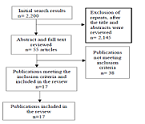 Strengths and Challenges to the Integration of Mental Health Services into the Primary Health Care System in Developing Countries: A Systematic ReviewAuthor: Roxanne Stowe MaloneyDOI: 10.21522/TIJAR.2014.09.02.Art001
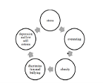 The Relationship between Adverse Life Experiences in Childhood and Unhealthy Eating Behaviour in Adulthood: A Literature ReviewAuthor: Marina Nagy Sabry SamyDOI: 10.21522/TIJAR.2014.09.02.Art003
 Does Gender Matter in the Adoption of Emails in the Namibian Enterprises?Author: Adalbertus KamanziDOI: 10.21522/TIJAR.2014.09.02.Art004
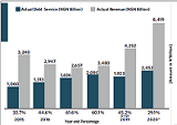 Exploring Nigeria Annual Budgets and Health Sector Budget Provisions towards Attainment of Universal Health Coverage Amid the Covid-19 Pandemic PreparednessAuthor: Gloria Nonyelum EnehDOI: 10.21522/TIJAR.2014.09.02.Art005
 An Investigation into Project Management Best Practices in Nigeria’s Telecommunication IndustryAuthor: Olanrewaju Modupe-SamuelDOI: 10.21522/TIJAR.2014.09.02.Art007
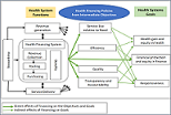 Perception Regarding Health Care Financing System and Its Advancement towards Universal Health Coverage in Nigeria among Residents of Awka, Anambra StateAuthor: Gloria Nonyelum EnehDOI: 10.21522/TIJAR.2014.09.02.Art008
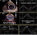 Normal Value Ranges of LV Deformation, Rotation and Twist Parameters in Healthy Adults by 4Dimensional XStrain Speckle Tracking EchocardiographyAuthor: Akhil MehrotraDOI: 10.21522/TIJAR.2014.09.02.Art009
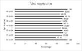 Factors Associated with Viral Load Suppression amongst People Living with HIV on Highly Active Anti-retroviral Therapy [Haart] at Kaloko, Chipulukusu and Kantolomba Art Health Facilities of Ndola DistrictAuthor: Nandipha HaneneDOI: 10.21522/TIJAR.2014.09.02.Art010
 Assessment of Knowledge and Acceptance of Covid-19 Vaccinations among Healthcare Workers in Kano State, NigeriaAuthor: Abdullahi AbduljaleelDOI: 10.21522/TIJAR.2014.09.02.Art011
 Assessing the Social and Economic Impact of Logistics Management on the Liberian Economy (The National Transit Authority 2015-2018)Author: Jeremiah Momo GbellayDOI: 10.21522/TIJAR.2014.09.02.Art012
 Information Overload and the Role of Librarians in Information Dissemination in Tertiary Institutions in Cross River State, NigeriaAuthor: Ebaye A.SDOI: 10.21522/TIJAR.2014.09.02.Art013
 Performance Appraisal System and its Implication on Employee Performance: A Study of Zambia Revenue AuthorityAuthor: Kausa Josephine KasongoDOI: 10.21522/TIJAR.2014.09.02.Art014
