References:
[1] Quninones MA, Greenberg BH, Kopelen HA, Koilpillai
C, Limacher MC, Shindler DM, et al. Echocardiographic predictors of clinical outcome
in patients with left ventricular dysfunction enrolled in the SOLVD registry and
trials: Significance of left ventricular hypertrophy, Studies of left ventricular
dysfunction. J Am CollCardiol2000; 35(5):1237-1244.
[2] Thune JJ. Kober L, Pfeffer MA, Skali H, Anavekar
NS, Bourgoun M, et al. Comparison of regional versus global assessment of left ventricular
function in patients with left ventricular dysfunction, heart failure, or both after
myocardial infarction; the valsartan in acute myocardial infarction echocardiographic
study. J Am Soc Echocardiogr 2006; 19(12):1462-1465.
[3] Kocabay G, Muraru D, Peluso D, Cucchini U, Mihaila
S, Padayattil-Jose Sanjay Pandey MD., et al. Normal left ventricular mechanics by
two-dimensional speckle-tracking echocardiography. Reference values I healthy adults.
Rev Esp Cardiol (Engl Ed)2014; 67(8):651-658.
[4] Quinones MA, Douglas PS, Foster E, Gorcsan J
3rd, Lewis JF, Pearlman AS, et al.; American Society of Echocardiography; Society
of Cardiovascular Anesthesiologists; Society of Pediatric Echocardiography. ACC/AHA
clinical competence statement on echocardiography: A report of the American College
of Cardiology/ American Heart Association/ American College of physicians-American
Society of Internal Medicine Task Force on Clinical Competence. J Am Soc-Echocardiogr.2003;
16(4):379-402.
[5] Nakatani S. Left ventricular and twist: Why
should we learn? J Cardiovasc Ultrasound 2011: 19(1): 1-6.
[6] Poveda F, Git D, Marti E, Andaluz A, Ballester
M, Carreras F, Helical Structure of the cardiac ventricular anatomy assessed by
diffusion tensor magnetic resonance imaging with multiresolution tractography. Rev
Esp Cardiol (Engl Ed)2013; 66(10): 782-790.
[7] Muraru, D.; Niero, A.; Zanella, H.R.; Cherata,
D.; Badano, L.P. Three-dimensional speckle-tracking echocardiography: Benefits and
limitation of integrating myocardial mechanics with three-dimensional imaging. Cardiovasc.
Diagn. Ther. 2018, 8, 101-117.
[8] Kang Y, Sun MM, Cui J, Chen HY, Su YG, Pan CZ,
et al. Three-dimensional speckle tracking echocardiography for the assessment of
left ventricular function and mechanical dyssynchrony. Acta Cardiol 2012; 67(4):423-430.
[9] Chen R, Wu X, Shen LJ, Wang B, Ma MM, Yang Y,
et al. Left ventricular myocardial function in hemodialysis and nondialysisuremia
patients: A three-dimensional speckle-tracking echocardiography study. PLoS One
2014; 9(6):e100265.
[10] Zhu M, Streiff C, Panosian J, Zhang Z, Song
X, Sahn DJ, et al. Regional Strain determination and myocardial infarction detection
by three-dimensional echocardiography with varied temporal resolution Echocardiography
2015; 32(2):339-348.
[11] Zhu M, Streiff C, Panosian J, Roundhill D, Lapin
M, Tutschek B, et al. Evaluation of stroke volume and ventricular mass in a fetal
heart model: A novel four-dimensional echocardiographic analysis. Echocardiography
2014; 31(9): 1138-1145.
[12] Jenkins C, Leano R, Chan J. Marwick TH. Reconstructed
versus real-time 3-dimensional echocardiography: Comparison with magnetic resonance
imaging. J Am Soc Echocardiogr 2007; 20(7): 862-868.
[13] Jenkins C, Leano R, Chan J, Marwick TH. Reconstructed
versus real-time 3-dimensional echocardiographic measurements of left ventricular
parameters using real-time three-dimensional echocardiography. J Am Coll Cardiol
2004; 44 (4):878-886.
[14] Pedrizzetti G, Mangual J and Tonti G. On the
geometrical relationship between global longitudinal strain and ejection fraction
in the evaluation of cardiac contraction. J Biomech. 2014 47:746-9.
[15] Stampehi MR, Mann DL, Nguyen JS, Cota F, Colmenares
C, and Dokainish H. Speckle strain echocardiography predicts outcome in patients
with heart failure with both depressed and preserved left ventricular ejection fraction.
Echocardiography. 2015; 32:71-8.
[16] Perk G, Tunick PA and Kronzon I. Non-Doppler
two-dimensional strain imaging by echocardiography from technical consideration
to clinical application. J Am Soc Echocardiogr.2007; 20:234-43.
[17] Mizuguchi Y, Oishi Y, Miyoshi H, luchi A, Nagase
N and Oki T. the functional role of longitudinal, circumferential, and radial myocardial
deformation for early impairment of left ventricular contraction and relaxation
in patients with cardiovascular risk factors: a study with two-dimensional strain
imaging. J Am Soc Echocardiogr. 2008; 21:1138-44.
[18] Share BL, La Gerche A, Naughton GA, Obert P,
and Kemp JG. Young Women with Abdominal Obesity Have Subclinical Myocardial Dysfunction.
Can J cardiol 2015; 31:1195-201.
[19] Yingchoncharoen T, Agarwal S, Popovic ZB, and
Marwick TH. Normal ranges of left ventricular strain: a meta-analysis. J Am Soc
Echocardiogr. 2013; 26:185-91.
[20] Kleijn SA, Pandian NG, Thomas JD, Perez de Isla
L, Kamp O, Zuber M, Nihoyannopoulos P, Forster T, Neser HJ, Geibel A, Gorissen W
and Zamorano JL. Normal reference values of left strain using three-dimensional
speckle tracking echocardiography: result from a multicenter study. Eur Heart J
Cardiovasc Imaging:2015; 16:410-6.
[21] Bernard A, Addetia K, Dulgheru R, Caballero
L, Sugimoto T, Akhaladze N, Athanasopoulos GD, Barone D, Baroni M, Cardim N, Hristova
K, Ilardi F, Lopez T, de la Morena G, Popescu BA, Penicka M, Ozyigit T, David Rodrigo
Carbonero J, van de Veire N, Stephan Von Bardeleben R, vinereanu D, Luis Zamorano
J, Martinez C, Magne J, Cosyns B, Donal E, Habib G, Badano LP, Lang RM and Lancellotti
P, 3D echocardiographic reference ranges for normal left ventricular and strain
results from the EACVI NORRE study. Eur Heart J Cardiovasc Imaging 2017; 18:475-483.
[22] Cheng S, Larson MG, McCabe EL, Osypiuk E, Lehman
BT, Stanchev P, Aragam J, Benjamin EJ, Solomon SD and Vasan RS. Age- and sex- based
reference limits and clinical correlates of myocardial strain and synchrony: the
Framingham Heart study. Circ Cardiovasc Imaging 2013; 6:692-9.
[23] Menting ME, mcGhie JS, Koopman LP, Vletter WB,
Helbing WA, van de Bosch AE and Roos-Hesselink JW. Normal myocardial strain values
using 2D speckle tracking echocardiography in healthy adults aged 20 to 72 years.
Echocardiography .2016;33:1665-1675.
[24] Dalen H, Thorstensen A, Aase SA, Ingul CB, Trop
H, Vatten LJ, and Stoylen A. Segmental and global longitudinal strain and strain
rate based on echocardiography of 1266 healthy individuals: the HUNT study in Norway.
Eur J Echocardiogr .2010; 11:176-83.
[25] Moreira HT, Nwabuo CC, Armstrong AC, Kishi S,
Gjesdal O, Reis JP. Schreiner PJ, Liu K, Lewis CE, Sidney S, Gidding SS, Lima JAC,
and Ambale-Venkatesh B. Reference Ranges and Regional Patterns of Left Ventricular
Strain and Strain Rate Using Two-Dimensional Speckle-Tracking Echocardiography in
a Healthy Middle-Aged Black and White Population: The CARDIA study. J Am Soc Echocardiogr.2017;30:647-658
e2.
[26] Park JH, Lee JH, Lee SY, Choi JO, Shin MS, Kim
MJ, Jung HO, Park JR, Sohn IS, Kim H, Park SM, Yoo NJ, Choi JH, Kim HK, Cho GY,
Lee MR, Park JS, Shim CY, Kim DH, Shin DH, Shin GJ, Shin SH, Kim KH, Kim WS, and
Park SW. Normal 2-Dimensional Strain Values of the Left Ventricular: A Substudy
of the Normal Echocardiographic Measurements in Korean Population Study. J Cardiovasc
Ultrasound.2016;24:285-293.
[27] Kaku K, Takeuchi M, Tsang W, Takigiku K, Yasukochi
S, Patel AR, Mor-Avi V, Lang RM and Otsuji Y. Age-related normal range of left ventricular
strain and torsion using three-dimensional speckle-tracking echocardiography. J
Am Soc Echocardiogr.2014;27:55-64.
[28] Liu CY, Lai S, Kawel-Boehm N, Chahal H, Ambale-Venkatesh
B, Lima JAC and Bluemke DA. Healthy aging of the left ventricle in relationship
to cardiovascular risk factors: The Multi-Ethnic Study of Atherosclerosis (MESA).
PLoS One.2017; 12: e0179947.
[29] Bjornstad P, Truong U, Pyls L, Dorosz JL, Cree-Green
M, Baumgartner A, Coe G, Regensteiner JG, Reusch JE and Nadeau KJ. Youth with type
1 diabetes have worse strain and less pronounced sex differences in early echocardiographic
markers of diabetic. Cardiomyopathy compared to their normoglycemic peers: A RESistance
to Insulin in Type 1 ANd Type 2 diabetes (RESISTANT) Study. J Diabetes Complications.
2016; 30:1103-10.
[30] Szelenyi Z, Fazakas A, Szenasi G, Tegze N, Fekete
B, Molvarec A, Hadusfalvy-Sudar S, Janosi O, Kiss M, Karadi I and Vereckei A. The
mechanism of reduced longitudinal left ventricular systolic function in hypertensive
patients with normal ejection fraction. J Hypertens. 2015; 33:1962-9; discussion
1969.
[31] Huang J, Yan ZN, Rui YF, Fan L, Shen D and Chen
DL. Left ventricular Systolic Function Changes in Primary Hypertension Patients
Detected by the Strain of Different Myocardium Layers. Medicine (Baltimore).2016;95:
e2440.
[32] Almeida AL, Teixido-Tura G, Choi EY, Opdahi
A, Fernandes VR, Wu CO, Bluemke DA and Lima JA. Metabolic syndrome, strain, and
reduced myocardial function: multi-ethnic study of atherosclerosis. Arq Bras Cardiol.
2014; 102:327-35.
[33] Pascual M, Pascual DA, Soria F, Vicente T, Hernandez
AM, Tebar FJ and Valdes M. Effects of isolated obesity on systolic and diastolic
left ventricular function. Heart. 2003; 89:1152-6.
[34] Friedewald WT, Levy RI, Fredrickson DS. Estimation
of the concentration of low densitylipoprotein in plasma, without use of the preparative
ultracentrifuge. Clin Chem. 1972; 18:499-502.
[35] Wagner M, Tiffe T, Morbach C, Gelbrich G, Stork
S, Heuschmann PU and Consortium S. characteristics and Course of Heart Failure Stages
A-B and Determinants of Progressiondesign and rationale of the STAAB cohort study.
Eur J Prev Cardiol. 2017; 24:468-479.
[36] Morbach C, Gelbrich G, at al. Impact of acquisition
and interpretation on total inter-observer variability in echocardiography: results
from the quality assurance program of the STAAB cohort study. Int J Cardiovasc Imaging.
2018.
[37] Bia D, Aguirre I, Zocalo Y, Devera L, Cabrera
Fischer E, Armentano R. [Regional differences in velocity, elasticity and wall buffering
function in systemic arteries: pulse wave analysis of the arterial pressure-diameter
relationship]. Rev Esp Cardiol. 2005; 58:167-74.
[38] Lehmann ED. Non invasive measurements of aortic
stiffness: methodological considerations. Pathol Biol. 1999; 47:716-30.
[39] Lantelme P, Mestre C, Lievre M, Gressard A,
Milon H. Heart rate AN important confounder of pulse wave velocity assessment Hypertension.
2002; 39:1083-7.
[40] Benetos A, Laurent S, Hoeks AP, Boutouyrie PH,
Safar ME. Arterial alterations with ageing and high blood pressure: a non-invasive
study of carotid and femoral arteries. Arterioscler Thromb. 1993; 13:90-7.
[41] Lang RM, Badano LP, Mor-Avi V et al (2015) Recommendations
for cardiac chamber quantification by echocardiography in adults: an update from
the American Society of Echocardiography and the European Association of Cardiovascular
Imaging. J Am Soc Echocardiogr 28:1-39.
[42] Ethnic-Specific Normative Reference Values for
Echocardiographic LA and, size LV (2015) Mass, and systolic functions:
the EchoNoRMAL study.JACC Cardiovas Imaging 8:656-665.
[43] Chahal NS, Lim TK, Jain P, Chmabers JC, Kooner
JS, Senior R (2010) Ethnicity-related differences in left ventricular functions,
structure, and geometry: a population study of UK Indian Asian and European white
subjects. Heart 96:466-471.
[44] Chahal NS, Lim TK, Jain P. Chmabers JC, Kooner
JS, Senior R (2012) Population-based references values for 3D echocardiographic
LV volumes and ejection fraction. JACC Cardiovasc Imaging 5:1191-1197.
[45] Bansal M, Mohan JC, Sengupta SP (2016) Normal
echocardiographic measurements in Indian adults: how different are we from the western
populations? A pilot study. Indian Heart J 68:772-775.
[46] Poppe KK, Doughty RN, Walsh HJ, Triggs CM, Whalley
GA (2014) A comparison of the effects of indexation on standard echocardiography
measurements of the left heart in a healthy multi-racial population. Int J Cardiovasc
Imaging 30:749-758.
[47] Asch FM, Miyoshi T, Addetia K et al (2019) Similarities
and differences in left ventricular size and function among races and nationalities:
results of the world alliances of echocardiography normal values study. J Am Soc
Echocardiogr 32:1396-1406.
[48] Chahal NS, Lim TK, Jain P, Chambers JC, Senior
R. Population-based reference values for 3D echocardiographic LV volumes and ejection
fraction JACC Cardiovasc Imaging. 2012; 5:1191-1197.
[49] Bansal M, Mohan JC, Sengupta SP. Normal echocardiographic
measurements in Indian adults: how different are we from the western populations?
A pilot study. Indian Heart J. 2016; 68:772-775.
[50] Luis SA, Yamada A, Khandheria BK, Speranza V,
Benjamin A, Ischenko M, et al. Use of three-dimensional speckle-tracking echocardiography
for quantitative assessment of global left ventricular function: A comparative study
of three-dimensional echocardiography. J Am Soc Echocardiogr 2014;27 (3):285-291.
[51] Brown J, Jenkins C, Marwick TH, Use of myocardial
strain to assess global left ventricular function: a comparison with cardiac magnetic
resonance and 3-dimensional echocardiography. Am Heart J. 2009; 157(1):102. e1-e5.
[52] Mignot A, Donal E, Zaroui A, Reant P, Saleem
A, Hamon C, et al. Global longitudinal strain as a major predictor of cardiac events
in patients with depressed left ventricular function: A multicenter study. J Am
Soc Echocardiogr 2010; 23(10):1019-1024.
[53] Cho GY, Marwick TH, Kim HS, Kim MK, Hong KS,
Oh DJ. Global2-dimensional strain as a new prognosticator in patients with heart
failure. J Am Coll Cardiol 2009; 54(7):618-624.
[54] Kearney LG, Lu K, Ord M, Patel SK, Profitis
K, Matalanis G, et al. Global longitudinal strain is a strong independent predictor
of all-cause mortality in patients with aortic stenosis. Eur Heart J Cardiovasc
Imaging 2012; 13(10):827-833.
[55] Dahi JS, Videbaek L, Poulsen MK, Rudbaek TR,
Pellikka PA, Moller JE, Global Strain in severe aortic valve stenosis: Relation
to clinical outcome after aortic valve replacement. Circ Cardiovasc Imaging 2012;5(5):613-620.
[56] Woo JS, Kim WS, Yu TK, Ha SJ, Kim SY, Bae JH
et al. Prognostic value of serial global longitudinal strain measured by two-dimensional
speckle tracking echocardiography in patients with ST-segment elevation myocardial
infarction. Am J Cardiol 2011; 108(3):340-347.
[57] Chen X, Xie H, Erkamp R, Kim K, Jia C, Rubin
JM, et al. 3-D correlation-based speckle tracking. Ultrason Imaging 2005; 27(1):21-36.
[58] Mor-Avi V, Lang RM, Badano LP, Belohlavek M,
Cardim NM, Derumeaux G, et al. Current evolving echocardiographic techniques for
the quantitative evaluation of cardiac mechanics: ASE/EAE consensus statement on
methodology and indications andorsed by the Japanese Society of Echocardiography.
J Am Soc Echocardiogr 2011;24(3):277-313.
[59] Perez de Isla L, Balcones DC, Fernandez-Golfin
C, Marcos-Alberca P, Almeria C Rodrigo JL et al. Three-dimensional-wall motion tracking;
a new and faster tool for myocardial strain assessment; comparison with two-dimensional-wall
motion tracking. J Am Soc Echocardiogr 2009; 22:325-30.
[60] Maffessanti F, Nesser HJ, Weinert L, Steringer-Mascherbauer
R, Niel J. Gorissen W et al. Quantitative evaluation of regional left ventricular
function using three-dimensional speckle Tracking echocardiography in patients with
and without heart disease. Am J cardiol 2009;1041755-62.
[61] Kleijn SA, Aly MF, Terwee CB, van Rossum AC,
Kamp O. Three-dimensional speckle tracking echocardiography for automatic assessment
of global and regional left ventricular function based on area strain. J Am Soc
Echocardiogr 2011; 24:314-21.
[62] Marwick TH, Leano RL, Brown J, Sun JP, Hoffmann
R, Lysyansky P et al. Myocardial strain measurements with 2-dimensional speckle-tracking
echocardiography: definition of normal range. JACC Cardiovasc Imaging 2009;
2:80-4.
[63] Maffessanti F, Nesser HJ, Weinert L, Steringer-Mascherbauer
R, Niel J, Gorissen W et al. Quantitative evaluation of regional left ventricular
function using three-dimensional speckle tracking echocardiography in patients with
and without heart disease. Am J Cardiol 2009; 104:1755-62.
[64] Saito K, Okura H, Watanabe N, Hayashida A, Obase
K, lmai K et al. Comprehensive evaluation of left ventricular strain using speckle
tracking echocardiography in normal adults: comparison of three-dimensional and
two-dimensional approaches. J Am Soc Echocardiogr 2009; 22:1025-30.
[65] Bernard A, Addettia K, Dulgheru R, Caballero
L, Sugimoto T, Akhaladze N et al. 3D echocardiographic reference ranges for normal
left ventricular volume and strain: results from the EACVI NORRE study. Eur Heart
J Cardiovasc Imaging 2017; 18:475-83.
[66] Kocabay G, Muraru D, Peluso D, Cucchini U, Mihaila
S, Padayattil-Jose S et al. Normal left ventricular mechanics by two-dimensional
speckle-tracking echocardiography. References values in healthy adults. Rev Esp
Cardiol (Engl Ed) 2014:67:651-8.
[67] Muraru D, Cucchini U, Mihaila S, Miglioranza
MH, Aruta P, Caralli G et al. Left ventricular myocardial strain by three-dimensional
speckle-tracking echocardiography in healthy subjects: reference values and analysis
of their physiologic and technical determinants. J Am Soc Echocardiogr 2014;
27:858-71.

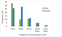 Effect of Duffy Antigen Receptor for Chemokines on Severity in Sickle Cell DiseaseAuthor: Aquel Rene LopezDOI: 10.21522/TIJAR.2014.09.01.Art001
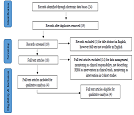 A Systematic Review to Observe the Impact of Risk-Based Monitoring as Compared to Conventional On-Site Monitoring in Randomised Clinical Trials and Quality Management in Large Cohort StudiesAuthor: Shubhra BansalDOI: 10.21522/TIJAR.2014.09.01.Art002
 Impact of Cultural Diversity on Overall Organizational Performance: A Moderating Role EducationAuthor: Laar David DiamDOI: 10.21522/TIJAR.2014.09.01.Art003
 Resettlement of Internally Displaced Persons in the North Central Geopolitical Zone of NigeriaAuthor: Askederin, F MDOI: 10.21522/TIJAR.2014.09.01.Art004
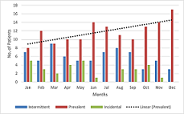 Integration of Mind and Skin; Psychological Co-morbidity in Dermatology and Skin Signs in PsychiatryAuthor: Bushra KhanDOI: 10.21522/TIJAR.2014.09.01.Art005
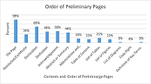 Basic Academic Research Structure and Format Guiding Principles for StudentsAuthor: Aquila Hakim M. JongroorDOI: 10.21522/TIJAR.2014.09.01.Art006
 An Assessment of Covid-19 Factors which Influence Non-Compliance of Payments in Respect of Social Security Contributions in GhanaAuthor: Samuel Nii Attoh AbbeyDOI: 10.21522/TIJAR.2014.09.01.Art007
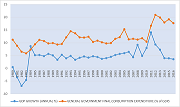 Government Expenditure on Economic Growth: Empirical Evidence from GhanaAuthor: Joshua AkanyongeDOI: 10.21522/TIJAR.2014.09.01.Art008
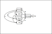 Impact of Human Resources Budgeting on Human Resource Management Accountability in Metropolitan, Municipal, and District Assemblies in the Ashanti RegionAuthor: Laar David DiamDOI: 10.21522/TIJAR.2014.09.01.Art009
 Tales of Early Childhood Education Teachers in Government Schools in Chipata, ZambiaAuthor: Daniel L. MpolomokaDOI: 10.21522/TIJAR.2014.09.01.Art010
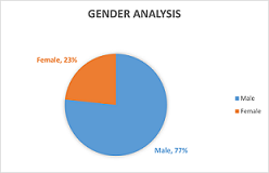 The Role Professional Accountant Firms play within the Liberian Market in Terms of Strategic Implementation of Financial Statement AuditAuthor: Jerome M. KessellyDOI: 10.21522/TIJAR.2014.09.01.Art011
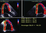 4Dimensional X Strain and 2Dimensional Speckle Tracking Echocardiographic Study: Normative Values of Strain Parameters of Left Ventricle and Tissue Doppler Imaging of Ascending Aorta in Healthy Adults –A Single Centre Indian StudyAuthor: Akhil MehrotraDOI: 10.21522/TIJAR.2014.09.01.Art012
