
Insertion of One-third Tubular Plate for Hemiepiphysiodesis in Children: Surgical Technique
Abstract:
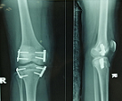
Limb length discrepancy and angular deformities are among the
main issues in paediatric patients. There are various ways to manage these two problems
which includes growth modulation with eight-Plate. For certain reasons, the use
of the eight-Plate is limited, thus conventional plates have been selected for the
guided growth, for example two-hole one-third tubular plate or reconstruction plate.
Literatures reported good results on its use but none of them explained the surgical
technique in details. Therefore, we proposed the surgical technique of inserting
the two-hole one-third tubular plate in hemiepiphysiodesis in children, which includes
pre-skin incision preparation, placement of one-third tubular plate and screw insertion.
The three steps are surgeon-friendly and less demanding, being suitable to apply
to paediatric patients that require growth modulation.
References:
[1] Jelinek,
E. M., Bittersohl, B., Martiny, F., Scharfstädt, A., Krauspe, R., & Westhoff,
B, 2012, The 8-plate versus physeal stapling for temporary hemiepiphyseodesis correcting
genu valgum and genu varum: a retrospective analysis of thirty five patients. International
orthopaedics, 36(3), 599–605. https://doi.org/10.1007/s00264-011-1369-5.
[2] Teresa
Cappello,2021, Expanded Indications for Guided Growth in Pediatric Extremities.
JPosna. Volume 3 (1), 1-14. https://doi.org/10.55275/JPOSNA-2021-217.
[3] Mycoskie
P. J., 1981, Complications of osteotomies about the knee in children. Orthopedic.
4(9), 1005–1015. https://doi.org/10.3928/0147-7447-19810901-04.
[4] Phemister,
D.B. (1933). Operative Arrestment of Longitudinial Growth of Bones in The Treatment
of Deformities, Journal of Bone and Joint Surgery. American Volume.
15, 1-15. Corpus ID: 74166208.
[5] Blount,
W.P, Clarke, 1949, Control of bone growth by epiphyseal stapling; a preliminary
report. The Journal of bone and joint surgery. American volume. 31A(3), 464–478.
PMID: 18153890.
[6] Campens,
C., Mousny, M., & Docquier, P. L., 2010, Comparison of three surgical epiphysiodesis
techniques for the treatment of lower limb length discrepancy. Acta orthopaedica
Belgica. 76(2), 226–232. PMID: 20503949.
[7] Nazareth,
A., Gyorfi, M. J., Rethlefsen, S. A., Wiseley, B., Noonan, K., & Kay, R. M ,2020,
Comparison of plate and screw constructs versus screws only for anterior distal
femoral hemiepiphysiodesis in children. Journal of pediatric orthopedics. Part
B, 29(1), 53–61. https://doi.org/10.1097/BPB.0000000000000661.
[8] Böhm, S.,
Krieg, A. H., Hefti, F., Brunner, R., Hasler, C. C., & Gaston, M. ,2013, Growth
guidance of angular lower limb deformities using a one-third two-hole tubular plate.
Journal of children's orthopaedics, 7(4), 289–294. https://doi.org/10.1007/s11832-013-0520-9.
[9] Stevens
P. M., 2007, Guided growth for angular correction: a preliminary series using a
tension band plate. Journal of pediatric orthopedics, 27(3), 253–259. https://doi.org/10.1097/BPO.0b013e31803433a1.
[10] Wiemann,
J. M., 4th, Tryon, C., & Szalay, E. A. ,2009, Physeal stapling versus 8-plate
hemiepiphysiodesis for guided correction of angular deformity about the knee.
Journal of pediatric orthopedics, 29(5), 481–485. https://doi.org/10.1097/BPO.0b013e3181aa24a8.
[11] Stevens P. M., 2023, Guided growth for FKFD: Historical
perspective and insights. Journal of children's orthopaedics, 17(3), 291. https://doi.org/10.1177/18632521231172994.
[12] Lebe M, van
Stralen RA, Buddhdev P. ,2022, Guided Growth of the Proximal Femur for the Management
of the 'Hip at Risk' in Children with Cerebral Palsy-A Systematic Review. Children
(Basel).;9(5):609. Published 2022 Apr 25. doi:10.3390/children9050609.
[13] Wang KK, Novacheck
TF, Rozumalski A, Georgiadis AG., 2019, Anterior Guided Growth of the Distal Femur
for Knee Flexion Contracture: Clinical, Radiographic, and Motion Analysis Results.
J Pediatr Orthop.; 39(5): e360-e365. doi:10.1097/BPO.0000000000001312.
[14] Bouchard M.,
2017, Guided Growth: Novel Applications in the Hip, Knee, and Ankle. J Pediatr
Orthop.;37 Suppl 2: S32-S36. doi:10.1097/BPO.0000000000001022.
[15] Metaizeau
JD, Denis D, Louis D., 2019, new femoral derotation technique based on guided growth
in children. Orthop Traumatol Surg Res.; 105(6):1175-1179. doi: 10.1016/j.otsr.2019.06.005.
[16] Danino B,
Rödl R, Herzenberg JE, et al., 2018, Guided growth: preliminary results of a multinational
study of 967 physes in 537 patients. J Child Orthop. 12(1):91-96. doi:10.1302/1863-2548.12.170050.
[17] Portinaro
N, Turati M, Cometto M, Bigoni M, Davids JR, Panou A. ,2019, Guided Growth of the
Proximal Femur for the Management of Hip Dysplasia in Children with Cerebral Palsy.
J Pediatr Orthop.;39(8): e622-e628. doi:10.1097/BPO.0000000000001069.
[18] Westberry
DE, Carpenter AM, Thomas JT, Graham GD, Pichiotino E, Hyer LC., 2020, Guided Growth
for Ankle Valgus Deformity: The Challenges of Hardware Removal. J Pediatr Orthop.;
40(9): e883-e888. doi:10.1097/BPO.0000000000001583.
[19] Gottliebsen
M, Shiguetomi-Medina JM, Rahbek O, Møller-Madsen B., 2016, Guided growth: mechanism
and reversibility of modulation. J Child Orthop.; 10(6):471-477. doi:10.1007/s11832-016-0778-9.
[20] Rethlefsen, S. A., Hanson, A. M., Wren, T. A. L., Abousamra, O., & Kay,
R. M. ,2020, Anterior distal femoral hemiepiphysiodesis with and without patellar
tendon shortening for fixed knee flexion contractures in children with cerebral
palsy. Journal of children's orthopaedics, 14(5), 415–420. https://doi.org/10.1302/1863-2548.14.200154.
